 Movie
Movie Controller

[English] 日本語
 Yorodumi
- PDB-2xf6: Crystal structure of Bacillus subtilis SPP1 phage gp23.1, a putat... -
+ Open data

- Basic information
| Entry | Database: PDB / ID: 2xf6 | ||||||
|---|---|---|---|---|---|---|---|
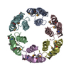
| Title | Crystal structure of Bacillus subtilis SPP1 phage gp23.1, a putative chaperone. | ||||||
 Components Components | GP23.1 | ||||||
 Keywords Keywords | VIRAL PROTEIN / CHAPERONE | ||||||
| Function / homology | Arc Repressor Mutant, subunit A - #1530 / : / SPP1 phage GP23.1 / Arc Repressor Mutant, subunit A / Orthogonal Bundle / Mainly Alpha / Bacteriophage SPP1 complete nucleotide sequence Function and homology information Function and homology information | ||||||
| Biological species |  BACILLUS PHAGE SPP1 (virus) BACILLUS PHAGE SPP1 (virus) | ||||||
| Method |  X-RAY DIFFRACTION / SYNCHROTRON / MOLECULAR REPLACEMENT / Resolution: 2.12 Å X-RAY DIFFRACTION / SYNCHROTRON / MOLECULAR REPLACEMENT / Resolution: 2.12 Å | ||||||
 Authors Authors | Veesler, D. / Blangy, S. / Lichiere, J. / Ortiz-Lombardia, M. / Tavares, P. / Campanacci, V. / Cambillau, C. | ||||||
 Citation Citation | Journal: Protein Sci. / Year: 2010 Title: Crystal Structure of Bacillus Subtilis Spp1 Phage Gp23.1, A Putative Chaperone. Authors: Veesler, D. / Blangy, S. / Lichiere, J. / Ortizlombardia, M. / Tavares, P. / Campanacci, V. / Cambillau, C. | ||||||
| History |
|
- Structure visualization
| Structure viewer | Molecule: MolmilJmol/JSmol |
|---|
- Downloads & links
-Download
| PDBx/mmCIF format | 2xf6.cif.gz | 33.1 KB | Display | PDBx/mmCIF format |
|---|---|---|---|---|
| PDB format | pdb2xf6.ent.gz | 22.9 KB | Display | PDB format |
| PDBx/mmJSON format | 2xf6.json.gz | Tree view | PDBx/mmJSON format | |
| Others |  Other downloads Other downloads |
-Validation report
| Arichive directory | https://data.pdbj.org/pub/pdb/validation_reports/xf/2xf6ftp://data.pdbj.org/pub/pdb/validation_reports/xf/2xf6 | HTTPS FTP |
|---|
-Related structure data


-Links
 PDBj
PDBj
- Assembly
| Deposited unit | 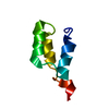
| ||||||||
|---|---|---|---|---|---|---|---|---|---|
| 1 | x 6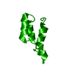
| ||||||||
| Unit cell |
| ||||||||
| Components on special symmetry positions |
|
-Components
| #1: Protein | Mass: 5761.278 Da / Num. of mol.: 1 Source method: isolated from a genetically manipulated source Source: (gene. exp.) BACILLUS PHAGE SPP1 (virus) / Plasmid: PETG20A / Production host:  |
|---|---|
| #2: Water | ChemComp-HOH /  Mass: 18.015 Da / Num. of mol.: 27 / Source method: isolated from a natural source / Formula: H2O Mass: 18.015 Da / Num. of mol.: 27 / Source method: isolated from a natural source / Formula: H2O |
-Experimental details
-Experiment
| Experiment | Method: X-RAY DIFFRACTION / Number of used crystals: 1 |
|---|
- Sample preparation
| Crystal | Density Matthews: 1.95 Å3/Da / Density % sol: 36.38 % / Description: NONE |
|---|---|
| Crystal grow | Details: 0.1 M NA HEPES PH7.5 20 PEG 10000 |
-Data collection
| Diffraction | Mean temperature: 100 K |
|---|---|
| Diffraction source | Source: SYNCHROTRON / Site: ESRF  / Beamline: BM14 / Wavelength: 1.7712 / Beamline: BM14 / Wavelength: 1.7712 |
| Detector | Type: MARRESEARCH / Detector: CCD / Date: Jun 17, 2009 |
| Radiation | Protocol: SINGLE WAVELENGTH / Monochromatic (M) / Laue (L): M / Scattering type: x-ray |
| Radiation wavelength | Wavelength: 1.7712 Å / Relative weight: 1 |
| Reflection | Resolution: 2.12→45.38 Å / Num. obs: 2826 / % possible obs: 99.7 % / Observed criterion σ(I): 2 / Redundancy: 11.2 % / Biso Wilson estimate: 21.89 Å2 / Rmerge(I) obs: 0.06 / Net I/σ(I): 33.7 |
| Reflection shell | Resolution: 2.12→2.24 Å / Redundancy: 8.3 % / Rmerge(I) obs: 0.24 / Mean I/σ(I) obs: 8.2 / % possible all: 98.2 |
- Processing
| Software |
| ||||||||||||||||||||||||||||||||||||||||||||||||||||||||||||||||||||||||||||||||||||||||||||||||||||||||||||||||||
|---|---|---|---|---|---|---|---|---|---|---|---|---|---|---|---|---|---|---|---|---|---|---|---|---|---|---|---|---|---|---|---|---|---|---|---|---|---|---|---|---|---|---|---|---|---|---|---|---|---|---|---|---|---|---|---|---|---|---|---|---|---|---|---|---|---|---|---|---|---|---|---|---|---|---|---|---|---|---|---|---|---|---|---|---|---|---|---|---|---|---|---|---|---|---|---|---|---|---|---|---|---|---|---|---|---|---|---|---|---|---|---|---|---|---|---|
| Refinement | Method to determine structure: MOLECULAR REPLACEMENT / Resolution: 2.12→45.38 Å / Cor.coef. Fo:Fc: 0.9091 / Cor.coef. Fo:Fc free: 0.8498 / Cross valid method: THROUGHOUT / σ(F): 0
| ||||||||||||||||||||||||||||||||||||||||||||||||||||||||||||||||||||||||||||||||||||||||||||||||||||||||||||||||||
| Displacement parameters | Biso mean: 29.59 Å2
| ||||||||||||||||||||||||||||||||||||||||||||||||||||||||||||||||||||||||||||||||||||||||||||||||||||||||||||||||||
| Refine analyze | Luzzati coordinate error obs: 0.302 Å | ||||||||||||||||||||||||||||||||||||||||||||||||||||||||||||||||||||||||||||||||||||||||||||||||||||||||||||||||||
| Refinement step | Cycle: LAST / Resolution: 2.12→45.38 Å
| ||||||||||||||||||||||||||||||||||||||||||||||||||||||||||||||||||||||||||||||||||||||||||||||||||||||||||||||||||
| Refine LS restraints |
| ||||||||||||||||||||||||||||||||||||||||||||||||||||||||||||||||||||||||||||||||||||||||||||||||||||||||||||||||||
| LS refinement shell | Resolution: 2.12→2.37 Å / Total num. of bins used: 5
| ||||||||||||||||||||||||||||||||||||||||||||||||||||||||||||||||||||||||||||||||||||||||||||||||||||||||||||||||||
| Refinement TLS params. | Method: refined / Refine-ID: X-RAY DIFFRACTION
| ||||||||||||||||||||||||||||||||||||||||||||||||||||||||||||||||||||||||||||||||||||||||||||||||||||||||||||||||||
| Refinement TLS group |
|
