Resolution: 1.6→40 Å / Num. obs: 17753 / % possible obs: 94.3 % / Observed criterion σ(I): 2 / Redundancy: 3.7 % / Biso Wilson estimate: 12.2 Å2 / Rmerge(I) obs: 0.08 / Net I/σ(I): 12.3
Reflection shell
Resolution: 1.6→1.64 Å / Redundancy: 3.5 % / Rmerge(I) obs: 0.39 / Mean I/σ(I) obs: 3.5 / % possible all: 80.2
-
Processing
Software
Name
Version
Classification
DENZO
datareduction
SCALEPACK
datascaling
SHELX
phasing
SHARP
phasing
REFMAC
5.2.0019
refinement
Refinement
Method to determine structure: SIRAS Starting model: NONE Resolution: 1.6→40 Å / Cor.coef. Fo:Fc: 0.957 / SU B: 1.319 / SU ML: 0.048 / Cross valid method: THROUGHOUT / ESU R: 0.102 / Stereochemistry target values: MAXIMUM LIKELIHOOD / Details: HYDROGENS HAVE BEEN ADDED IN THE RIDING POSITIONS.
Rfactor
Num. reflection
% reflection
Selection details
Rfree
0.209
915
5.2 %
RANDOM
Rwork
0.167
-
-
-
obs
0.1678
17742
93.93 %
-
Solvent computation
Ion probe radii: 0.8 Å / Shrinkage radii: 0.8 Å / VDW probe radii: 1.4 Å / Solvent model: MASK
 Movie
Movie Controller

 Yorodumi
 Open data
 Basic information
 Components
 Keywords
 Function and homology information
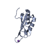
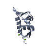
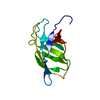
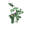
 YERSINIA PESTIS (bacteria)
 X-RAY DIFFRACTION /
  Authors
 Citation
 Structure visualization
 Downloads & links
 Other downloads


 PDBj
PDBj Assembly


 Mass: 96.063 Da / Num. of mol.: 3 / Source method: obtained synthetically / Formula: SO4
 Mass: 18.015 Da / Num. of mol.: 222 / Source method: isolated from a natural source / Formula: H2O
 Sample preparation
 Processing