 Movie
Movie Controller

[English] 日本語
 Yorodumi
- PDB-2xbk: X-ray structure of the substrate-bound cytochrome P450 PimD - a p... -
+ Open data

- Basic information
| Entry | Database: PDB / ID: 2xbk | ||||||
|---|---|---|---|---|---|---|---|
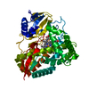
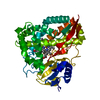
| Title | X-ray structure of the substrate-bound cytochrome P450 PimD - a polyene macrolide antibiotic pimaricin epoxidase | ||||||
 Components Components | PIMD PROTEIN | ||||||
 Keywords Keywords | OXIDOREDUCTASE / EPOXIDATION | ||||||
| Function / homology |  Function and homology information Function and homology informationoxidoreductase activity, acting on paired donors, with incorporation or reduction of molecular oxygen / monooxygenase activity / iron ion binding / heme binding Similarity search - Function | ||||||
| Biological species |  STREPTOMYCES NATALENSIS (bacteria) STREPTOMYCES NATALENSIS (bacteria) | ||||||
| Method |  X-RAY DIFFRACTION / SYNCHROTRON / MOLECULAR REPLACEMENT / Resolution: 1.95 Å X-RAY DIFFRACTION / SYNCHROTRON / MOLECULAR REPLACEMENT / Resolution: 1.95 Å | ||||||
 Authors Authors | Kells, P.M. / Ouellet, H. / Santos-Aberturas, J. / Aparicio, J.F. / Podust, L.M. | ||||||
 Citation Citation | Journal: Chem.Biol. / Year: 2010 Title: Structure of Cytochrome P450 Pimd Suggests Epoxidation of the Polyene Macrolide Pimaricin Occurs Via a Hydroperoxoferric Intermediate. Authors: Kells, P.M. / Ouellet, H. / Santos-Aberturas, J. / Aparicio, J.F. / Podust, L.M. | ||||||
| History |
|
- Structure visualization
| Structure viewer | Molecule: MolmilJmol/JSmol |
|---|
- Downloads & links
-Download
| PDBx/mmCIF format | 2xbk.cif.gz | 188.2 KB | Display | PDBx/mmCIF format |
|---|---|---|---|---|
| PDB format | pdb2xbk.ent.gz | 147.2 KB | Display | PDB format |
| PDBx/mmJSON format | 2xbk.json.gz | Tree view | PDBx/mmJSON format | |
| Others |  Other downloads Other downloads |
-Validation report
| Arichive directory | https://data.pdbj.org/pub/pdb/validation_reports/xb/2xbkftp://data.pdbj.org/pub/pdb/validation_reports/xb/2xbk | HTTPS FTP |
|---|
-Related structure data
| Related structure data |  2x9pSC S: Starting model for refinement C: citing same article ( |
|---|---|
| Similar structure data |


-Links
 PDBj
PDBj
- Assembly
| Deposited unit | 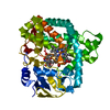
| ||||||||
|---|---|---|---|---|---|---|---|---|---|
| 1 |
| ||||||||
| Unit cell |
|
-Components
| #1: Protein | Mass: 44528.770 Da / Num. of mol.: 1 / Fragment: RESIDUES 5-397 Source method: isolated from a genetically manipulated source Details: RESIDUES UPSTREAM OF SER 5 INCLUDING 6XHIS TAG ARE ENGINEERED Source: (gene. exp.) STREPTOMYCES NATALENSIS (bacteria) / Plasmid: PQE-30 / Production host: |
|---|---|
| #2: Chemical | ChemComp-HEM /   Mass: 616.487 Da / Num. of mol.: 1 / Source method: obtained synthetically / Formula: C34H32FeN4O4 Mass: 616.487 Da / Num. of mol.: 1 / Source method: obtained synthetically / Formula: C34H32FeN4O4 |
| #3: Chemical | ChemComp-XBK /   Mass: 649.726 Da / Num. of mol.: 1 / Source method: obtained synthetically / Formula: C33H47NO12 Mass: 649.726 Da / Num. of mol.: 1 / Source method: obtained synthetically / Formula: C33H47NO12 |
| #4: Water | ChemComp-HOH /  Mass: 18.015 Da / Num. of mol.: 272 / Source method: isolated from a natural source / Formula: H2O Mass: 18.015 Da / Num. of mol.: 272 / Source method: isolated from a natural source / Formula: H2O |
| Nonpolymer details | 4,5-DEEPOXYPIMARICIN: NON-EPOXIDATED PRECURSOR OF THE POLYENE ANTIBIOTIC PIMARICIN OR NATAMYCIN. ...4,5-DEEPOXYPIM |
| Sequence details | 6XHIS TAG ENGINEERED |
-Experimental details
-Experiment
| Experiment | Method: X-RAY DIFFRACTION / Number of used crystals: 1 |
|---|
- Sample preparation
| Crystal | Density Matthews: 2.87 Å3/Da / Density % sol: 57.2 % / Description: NONE |
|---|---|
| Crystal grow | Temperature: 296 K / pH: 7 Details: 0.2 M SODIUM MALONATE, PH 7.0; 20% PEG 3330, T=23 C |
-Data collection
| Diffraction | Mean temperature: 110 K |
|---|---|
| Diffraction source | Source: SYNCHROTRON / Site: ALS  / Beamline: 8.3.1 / Wavelength: 1.11587 / Beamline: 8.3.1 / Wavelength: 1.11587 |
| Detector | Type: ADSC QUANTUM 315 / Detector: CCD / Date: Jul 15, 2009 / Details: MIRRORS |
| Radiation | Monochromator: SI (111) DOUBLE CRYSTAL / Protocol: SINGLE WAVELENGTH / Monochromatic (M) / Laue (L): M / Scattering type: x-ray |
| Radiation wavelength | Wavelength: 1.11587 Å / Relative weight: 1 |
| Reflection | Resolution: 1.95→17.01 Å / Num. obs: 38650 / % possible obs: 99.8 % / Observed criterion σ(I): 1.5 / Redundancy: 4.1 % / Biso Wilson estimate: 30.1 Å2 / Rmerge(I) obs: 0.07 / Net I/σ(I): 10 |
| Reflection shell | Resolution: 1.95→2.06 Å / Redundancy: 4.1 % / Rmerge(I) obs: 0.52 / Mean I/σ(I) obs: 2.3 / % possible all: 100 |
- Processing
| Software |
| ||||||||||||||||||||||||||||||||||||||||||||||||||||||||||||||||||||||||||||||||||||||||||||||||||||||||||||||||||||||||||||||||||||||||||||||||||||||||||||||||||||||||||||||||||||||
|---|---|---|---|---|---|---|---|---|---|---|---|---|---|---|---|---|---|---|---|---|---|---|---|---|---|---|---|---|---|---|---|---|---|---|---|---|---|---|---|---|---|---|---|---|---|---|---|---|---|---|---|---|---|---|---|---|---|---|---|---|---|---|---|---|---|---|---|---|---|---|---|---|---|---|---|---|---|---|---|---|---|---|---|---|---|---|---|---|---|---|---|---|---|---|---|---|---|---|---|---|---|---|---|---|---|---|---|---|---|---|---|---|---|---|---|---|---|---|---|---|---|---|---|---|---|---|---|---|---|---|---|---|---|---|---|---|---|---|---|---|---|---|---|---|---|---|---|---|---|---|---|---|---|---|---|---|---|---|---|---|---|---|---|---|---|---|---|---|---|---|---|---|---|---|---|---|---|---|---|---|---|---|---|
| Refinement | Method to determine structure: MOLECULAR REPLACEMENT Starting model: PDB ENTRY 2X9P Resolution: 1.95→67.88 Å / Cor.coef. Fo:Fc: 0.972 / Cor.coef. Fo:Fc free: 0.95 / SU B: 7.439 / SU ML: 0.094 / Cross valid method: THROUGHOUT / ESU R: 0.245 / ESU R Free: 0.131 / Stereochemistry target values: MAXIMUM LIKELIHOOD Details: HYDROGENS HAVE BEEN ADDED IN THE RIDING POSITIONS. RESIDUES WITH THE SIDE CHAIN MISSING FROM THE ELECTRON DENSITY WERE MODELED AS ALANINE
| ||||||||||||||||||||||||||||||||||||||||||||||||||||||||||||||||||||||||||||||||||||||||||||||||||||||||||||||||||||||||||||||||||||||||||||||||||||||||||||||||||||||||||||||||||||||
| Solvent computation | Ion probe radii: 0.8 Å / Shrinkage radii: 0.8 Å / VDW probe radii: 1.4 Å / Solvent model: MASK | ||||||||||||||||||||||||||||||||||||||||||||||||||||||||||||||||||||||||||||||||||||||||||||||||||||||||||||||||||||||||||||||||||||||||||||||||||||||||||||||||||||||||||||||||||||||
| Displacement parameters | Biso mean: 30.816 Å2
| ||||||||||||||||||||||||||||||||||||||||||||||||||||||||||||||||||||||||||||||||||||||||||||||||||||||||||||||||||||||||||||||||||||||||||||||||||||||||||||||||||||||||||||||||||||||
| Refinement step | Cycle: LAST / Resolution: 1.95→67.88 Å
| ||||||||||||||||||||||||||||||||||||||||||||||||||||||||||||||||||||||||||||||||||||||||||||||||||||||||||||||||||||||||||||||||||||||||||||||||||||||||||||||||||||||||||||||||||||||
| Refine LS restraints |
|