- PDB-2xb0: DNA-binding domain from Saccharomyces cerevisiae chromatin- remod... -
+
Open data
ID or keywords:
Loading...
-
Basic information
Entry
Database: PDB / ID: 2xb0
Title
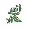
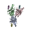
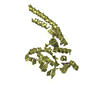
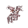
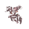
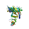
DNA-binding domain from Saccharomyces cerevisiae chromatin- remodelling protein Chd1
Components
CHROMO DOMAIN-CONTAINING PROTEIN 1
Keywords
HYDROLASE / DNA-BINDING PROTEIN / TRANSCRIPTION / CHROMATIN REGULATOR
Function / homology
Function and homology information
regulation of transcriptional start site selection at RNA polymerase II promoter / nucleolar chromatin / negative regulation of DNA-templated DNA replication / regulation of chromatin organization / SLIK (SAGA-like) complex / DNA double-strand break processing / rDNA binding / nucleosome organization / nucleosome array spacer activity / histone H3K4me3 reader activity ...regulation of transcriptional start site selection at RNA polymerase II promoter / nucleolar chromatin / negative regulation of DNA-templated DNA replication / regulation of chromatin organization / SLIK (SAGA-like) complex / DNA double-strand break processing / rDNA binding / nucleosome organization / nucleosome array spacer activity / histone H3K4me3 reader activity / SAGA complex / ATP-dependent chromatin remodeler activity / sister chromatid cohesion / termination of RNA polymerase II transcription / termination of RNA polymerase I transcription / ATP-dependent activity, acting on DNA / transcription elongation by RNA polymerase II / chromatin DNA binding / double-strand break repair via homologous recombination / Hydrolases; Acting on acid anhydrides; Acting on acid anhydrides to facilitate cellular and subcellular movement / site of double-strand break / histone binding / transcription cis-regulatory region binding / chromatin remodeling / chromatin binding / regulation of transcription by RNA polymerase II / chromatin / ATP hydrolysis activity / mitochondrion / DNA binding / ATP binding / nucleus Similarity search - Function
Mass: 18.015 Da / Num. of mol.: 109 / Source method: isolated from a natural source / Formula: H2O
Sequence details
PARTIAL SEQUENCE (AA 1009-1274) CORRESPONDING TO THE DNA- BINDING DOMAIN OF YEAST CHD1. FIRST FOUR ...PARTIAL SEQUENCE (AA 1009-1274) CORRESPONDING TO THE DNA- BINDING DOMAIN OF YEAST CHD1. FIRST FOUR RESIDUES (GPLG) ARE NON-NATIVE AND ARE LEFT FROM CLEAVAGE OF THE GST-TAG WITH PRESCISSION PROTEASE.
-
Experimental details
-
Experiment
Experiment
Method: X-RAY DIFFRACTION / Number of used crystals: 1
-
Sample preparation
Crystal
Density Matthews: 2.77 Å3/Da / Density % sol: 55.57 % / Description: NONE
Crystal grow
pH: 7.5 / Details: 0.1 M TRIS, PH 8.5, 28% PEG 4K, 0.35 M MGCL2
Resolution: 2→49.2 Å / Num. obs: 22943 / % possible obs: 97.5 % / Observed criterion σ(I): 2 / Redundancy: 9.7 % / Biso Wilson estimate: 34.64 Å2 / Rmerge(I) obs: 0.08 / Net I/σ(I): 18.1
Reflection shell
Resolution: 2→2.11 Å / Redundancy: 9.4 % / Rmerge(I) obs: 0.82 / Mean I/σ(I) obs: 2.5 / % possible all: 95.9
-
Processing
Software
Name
Version
Classification
PHENIX
(PHENIX.REFINE)
refinement
MOSFLM
datareduction
SCALA
datascaling
PHENIX
phasing
Refinement
Method to determine structure: MAD Starting model: NONE Resolution: 2→49.255 Å / SU ML: 0.28 / σ(F): 1.34 / Phase error: 25.5 / Stereochemistry target values: ML Details: RIDING HYDROGENS WERE USED IN REFINEMENT AMINO ACIDS 209-240 (NEVHNPVAKKSASSSDTTPTPSKKGKGITGSS) AND 266-270 (TKSPS) ARE DISORDERED WERE NOT MODELLED OWING TO INSUFFICIENT ELECTRON DENSITY.
Rfactor
Num. reflection
% reflection
Rfree
0.2479
2263
9.9 %
Rwork
0.205
-
-
obs
0.2093
22894
97.18 %
Solvent computation
Shrinkage radii: 0.9 Å / VDW probe radii: 1.11 Å / Solvent model: FLAT BULK SOLVENT MODEL / Bsol: 66.114 Å2 / ksol: 0.399 e/Å3
Displacement parameters
Biso mean: 61.3 Å2
Baniso -1
Baniso -2
Baniso -3
1-
1.8428 Å2
0 Å2
0 Å2
2-
-
5.0238 Å2
0 Å2
3-
-
-
-6.8666 Å2
Refinement step
Cycle: LAST / Resolution: 2→49.255 Å
Protein
Nucleic acid
Ligand
Solvent
Total
Num. atoms
1842
0
14
109
1965
Refine LS restraints
Refine-ID
Type
Dev ideal
Number
X-RAY DIFFRACTION
f_bond_d
0.018
1938
X-RAY DIFFRACTION
f_angle_d
1.359
2598
X-RAY DIFFRACTION
f_dihedral_angle_d
14.263
752
X-RAY DIFFRACTION
f_chiral_restr
0.085
279
X-RAY DIFFRACTION
f_plane_restr
0.005
330
LS refinement shell
Resolution (Å)
Rfactor Rfree
Num. reflection Rfree
Rfactor Rwork
Num. reflection Rwork
Refine-ID
% reflection obs (%)
2-2.0715
0.3591
208
0.327
1989
X-RAY DIFFRACTION
95
2.0715-2.1544
0.3008
208
0.2838
2026
X-RAY DIFFRACTION
98
2.1544-2.2525
0.3087
210
0.2413
2029
X-RAY DIFFRACTION
96
2.2525-2.3713
0.2817
233
0.2126
2002
X-RAY DIFFRACTION
96
2.3713-2.5198
0.2563
206
0.1984
2062
X-RAY DIFFRACTION
97
2.5198-2.7144
0.2583
248
0.1961
2021
X-RAY DIFFRACTION
98
2.7144-2.9875
0.2676
228
0.2091
2084
X-RAY DIFFRACTION
98
2.9875-3.4197
0.2662
246
0.2173
2072
X-RAY DIFFRACTION
98
3.4197-4.3081
0.2323
234
0.1754
2120
X-RAY DIFFRACTION
99
4.3081-49.2699
0.1995
242
0.1866
2226
X-RAY DIFFRACTION
97
Refinement TLS params.
Method: refined / Refine-ID: X-RAY DIFFRACTION
ID
L11 (°2)
L12 (°2)
L13 (°2)
L22 (°2)
L23 (°2)
L33 (°2)
S11 (Å °)
S12 (Å °)
S13 (Å °)
S21 (Å °)
S22 (Å °)
S23 (Å °)
S31 (Å °)
S32 (Å °)
S33 (Å °)
T11 (Å2)
T12 (Å2)
T13 (Å2)
T22 (Å2)
T23 (Å2)
T33 (Å2)
Origin x (Å)
Origin y (Å)
Origin z (Å)
1
0.5134
0.261
0.404
2.4028
2.0332
1.824
-0.0189
-0.1516
-0.0188
0.7428
0.1675
-0.2586
0.33
0.2885
-0.1368
0.4699
-0.0347
-0.0893
0.321
0.0324
0.2512
10.6756
65.3063
79.2636
2
2.3943
1.512
0.9081
3.2675
0.0964
0.8162
-0.2821
0.2469
-0.0041
-1.0554
0.3969
-0.0605
0.1143
0.0501
-0.0829
0.8428
-0.0351
-0.0412
0.3014
-0.0089
0.2257
-10.6744
34.2717
55.7702
3
2.3422
0.1727
2.3681
0.718
1.5019
5.1544
0.2534
-0.1417
-0.7285
0.0753
-0.6913
-0.1426
0.8482
-0.0586
0.1162
0.6494
-0.1447
-0.0506
0.3681
0.1101
0.3873
-1.2847
52.3863
74.4755
4
1.9885
0.9547
0.3068
1.3934
0.8226
1.8499
0.0822
-0.1246
0.632
0.0012
-0.1703
0.0882
-0.0359
-0.2246
0.1405
0.063
0.0027
-0.0073
0.1728
-0.1003
0.271
-1.831
75.6372
58.5856
5
0.9192
0.722
0.5578
0.6396
0.4402
0.406
0.3516
-1.4781
0.9828
0.3615
-0.8365
0.4929
0.1704
-0.7768
0.4399
0.1251
-0.166
0.0986
0.8782
-0.4324
0.5123
-15.494
75.1158
66.0314
6
1.6454
1.1824
-1.5309
2.2534
0.6436
3.7066
0.1182
-0.1278
-0.1652
0.3804
-0.3762
-0.2347
0.6517
-0.8418
0.2
0.1906
-0.1045
-0.0073
0.2784
-0.0336
0.2942
-4.3266
63.979
56.8805
Refinement TLS group
ID
Refine-ID
Refine TLS-ID
Selection details
1
X-RAY DIFFRACTION
1
(CHAINXANDRESID1:60)
2
X-RAY DIFFRACTION
2
(CHAINXANDRESID61:108)
3
X-RAY DIFFRACTION
3
(CHAINXANDRESID109:128)
4
X-RAY DIFFRACTION
4
(CHAINXANDRESID129:205)
5
X-RAY DIFFRACTION
5
(CHAINXANDRESID206:250)
6
X-RAY DIFFRACTION
6
(CHAINXANDRESID251:266)
+
About Yorodumi
-
News
-
Feb 9, 2022. New format data for meta-information of EMDB entries
New format data for meta-information of EMDB entries
Version 3 of the EMDB header file is now the official format.
The previous official version 1.9 will be removed from the archive.
In the structure databanks used in Yorodumi, some data are registered as the other names, "COVID-19 virus" and "2019-nCoV". Here are the details of the virus and the list of structure data.
Jan 31, 2019. EMDB accession codes are about to change! (news from PDBe EMDB page)
EMDB accession codes are about to change! (news from PDBe EMDB page)
The allocation of 4 digits for EMDB accession codes will soon come to an end. Whilst these codes will remain in use, new EMDB accession codes will include an additional digit and will expand incrementally as the available range of codes is exhausted. The current 4-digit format prefixed with “EMD-” (i.e. EMD-XXXX) will advance to a 5-digit format (i.e. EMD-XXXXX), and so on. It is currently estimated that the 4-digit codes will be depleted around Spring 2019, at which point the 5-digit format will come into force.
The EM Navigator/Yorodumi systems omit the EMD- prefix.
Related info.:Q: What is EMD? / ID/Accession-code notation in Yorodumi/EM Navigator
Yorodumi is a browser for structure data from EMDB, PDB, SASBDB, etc.
This page is also the successor to EM Navigator detail page, and also detail information page/front-end page for Omokage search.
The word "yorodu" (or yorozu) is an old Japanese word meaning "ten thousand". "mi" (miru) is to see.
Related info.:EMDB / PDB / SASBDB / Comparison of 3 databanks / Yorodumi Search / Aug 31, 2016. New EM Navigator & Yorodumi / Yorodumi Papers / Jmol/JSmol / Function and homology information / Changes in new EM Navigator and Yorodumi
 Movie
Movie Controller

 Yorodumi
 Open data
 Basic information
 Components
 Keywords
 Function and homology information
 X-RAY DIFFRACTION /
  Authors
 Citation
 Structure visualization
 Downloads & links
 Other downloads


 PDBj
PDBj Assembly
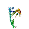


 Mass: 92.094 Da / Num. of mol.: 2 / Source method: obtained synthetically / Formula: C3H8O3
 Mass: 35.453 Da / Num. of mol.: 2 / Source method: obtained synthetically / Formula: Cl
 Mass: 18.015 Da / Num. of mol.: 109 / Source method: isolated from a natural source / Formula: H2O
 Sample preparation
 / Beamline: I04 / Wavelength: 0.9775
  Processing