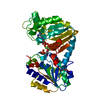
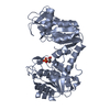
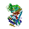
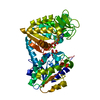
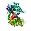
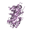
[English] 日本語
Yorodumi
- PDB-2wzd: The catalytically active fully closed conformation of human phosp... -

+
Open data


ID or keywords:

Loading...

-
Basic information

Entry
Database: PDB / ID: 2wzd
TitleThe catalytically active fully closed conformation of human phosphoglycerate kinase K219A mutant in complex with ADP, 3PG and aluminium trifluoride
ComponentsPHOSPHOGLYCERATE KINASE 1
KeywordsTRANSFERASE / HEREDITARY HEMOLYTIC ANEMIA / PHOSPHOPROTEIN / KINASE / GLYCOLYSIS / NUCLEOTIDE-BINDING
Function / homology
Function and homology information


Manipulation of host energy metabolism / phosphoglycerate kinase / phosphoglycerate kinase activity / protein-disulfide reductase (NAD(P)H) activity / Gluconeogenesis / canonical glycolysis / Glycolysis / plasminogen activation / epithelial cell differentiation / negative regulation of angiogenesis ...Manipulation of host energy metabolism / phosphoglycerate kinase / phosphoglycerate kinase activity / protein-disulfide reductase (NAD(P)H) activity / Gluconeogenesis / canonical glycolysis / Glycolysis / plasminogen activation / epithelial cell differentiation / negative regulation of angiogenesis / ADP binding / gluconeogenesis / glycolytic process / cellular response to hypoxia / membrane raft / phosphorylation / extracellular space / extracellular exosome / ATP binding / membrane / cytosol
Similarity search - Function
Phosphoglycerate kinase, N-terminal domain / Phosphoglycerate kinase / Phosphoglycerate kinase, N-terminal / Phosphoglycerate kinase, conserved site / Phosphoglycerate kinase superfamily / Phosphoglycerate kinase / Phosphoglycerate kinase signature. / Rossmann fold / 3-Layer(aba) Sandwich / Alpha Beta
Similarity search - Domain/homology
3-PHOSPHOGLYCERIC ACID / ADENOSINE-5'-DIPHOSPHATE / ALUMINUM FLUORIDE / Phosphoglycerate kinase 1
Similarity search - Component
Biological speciesHOMO SAPIENS (human)
MethodX-RAY DIFFRACTION / SYNCHROTRON / MOLECULAR REPLACEMENT / Resolution: 1.56 Å
AuthorsBowler, M.W. / Cliff, M.J. / Marston, J.P.M. / Baxter, N.J. / Hownslow, A.M.H. / Varga, A.V. / Szabo, J. / Vas, M. / Blackburn, G.M. / Waltho, J.P.
CitationJournal: J.Am.Chem.Soc. / Year: 2010
Title: Transition State Analogue Structures of Human Phosphoglycerate Kinase Establish the Importance of Charge Balance in Catalysis.
Authors: Cliff, M.J. / Bowler, M.W. / Szabo, J. / Marston, J.P.M. / Varga, A.V. / Hownslow, A.M.H. / Baxter, N.J. / Blackburn, G.M. / Vas, M. / Waltho, J.P.
History
DepositionNov 27, 2009Deposition site: PDBE / Processing site: PDBE
Revision 1.0Apr 14, 2010Provider: repository / Type: Initial release
Revision 1.1Jul 13, 2011Group: Advisory / Version format compliance
Revision 1.2Dec 7, 2011Group: Database references
Revision 1.3Dec 20, 2023Group: Data collection / Database references ...Data collection / Database references / Derived calculations / Other / Refinement description
Category: chem_comp_atom / chem_comp_bond ...chem_comp_atom / chem_comp_bond / database_2 / pdbx_database_status / pdbx_initial_refinement_model / pdbx_struct_conn_angle / struct_conn / struct_site
Item: _database_2.pdbx_DOI / _database_2.pdbx_database_accession ..._database_2.pdbx_DOI / _database_2.pdbx_database_accession / _pdbx_database_status.status_code_sf / _pdbx_struct_conn_angle.ptnr1_auth_comp_id / _pdbx_struct_conn_angle.ptnr1_auth_seq_id / _pdbx_struct_conn_angle.ptnr1_label_asym_id / _pdbx_struct_conn_angle.ptnr1_label_atom_id / _pdbx_struct_conn_angle.ptnr1_label_comp_id / _pdbx_struct_conn_angle.ptnr3_auth_comp_id / _pdbx_struct_conn_angle.ptnr3_auth_seq_id / _pdbx_struct_conn_angle.ptnr3_label_asym_id / _pdbx_struct_conn_angle.ptnr3_label_atom_id / _pdbx_struct_conn_angle.ptnr3_label_comp_id / _pdbx_struct_conn_angle.value / _struct_conn.pdbx_dist_value / _struct_conn.ptnr1_auth_comp_id / _struct_conn.ptnr1_auth_seq_id / _struct_conn.ptnr1_label_asym_id / _struct_conn.ptnr1_label_atom_id / _struct_conn.ptnr1_label_comp_id / _struct_conn.ptnr1_label_seq_id / _struct_conn.ptnr2_auth_comp_id / _struct_conn.ptnr2_auth_seq_id / _struct_conn.ptnr2_label_asym_id / _struct_conn.ptnr2_label_atom_id / _struct_conn.ptnr2_label_comp_id / _struct_conn.ptnr2_label_seq_id / _struct_site.pdbx_auth_asym_id / _struct_site.pdbx_auth_comp_id / _struct_site.pdbx_auth_seq_id

-
Structure visualization

Structure viewerMolecule:
MolmilJmol/JSmol

Downloads & links

-
Assembly

Deposited unit
A: PHOSPHOGLYCERATE KINASE 1
hetero molecules


Theoretical massNumber of molelcules
Total (without water)45,3726
Polymers44,6151
Non-polymers7575
Water8,395466
1


  • Idetical with deposited unit
  • defined by author&software
TypeNameSymmetry operationNumber
identity operation1_555x,y,z1
MethodPISA
Unit cell
Length a, b, c (Å)39.380, 91.590, 108.480
Angle α, β, γ (deg.)90.00, 90.00, 90.00
Int Tables number19
Space group name H-MP212121

-
Components

-
Protein , 1 types, 1 molecules A

#1: Protein PHOSPHOGLYCERATE KINASE 1 / / PRIMER RECOGNITION PROTEIN 2 / PRP 2 / CELL MIGRATION-INDUCING GENE 10 PROTEIN


Mass: 44614.520 Da / Num. of mol.: 1 / Fragment: RESIDUES 2-417 / Mutation: YES
Source method: isolated from a genetically manipulated source
Details: COMPLEXED WITH ADP, 3PG AND ALUMINIUM TRIFLUORIDE / Source: (gene. exp.) HOMO SAPIENS (human) / Production host: ESCHERICHIA COLI (E. coli) / Strain (production host): BL21 / References: UniProt: P00558, phosphoglycerate kinase

-
Non-polymers , 6 types, 471 molecules

#2: Chemical ChemComp-MG / MAGNESIUM ION


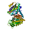
Mass: 24.305 Da / Num. of mol.: 1 / Source method: obtained synthetically / Formula: Mg
#3: Chemical ChemComp-CL / CHLORIDE ION / Chloride


Mass: 35.453 Da / Num. of mol.: 1 / Source method: obtained synthetically / Formula: Cl
#4: Chemical ChemComp-AF3 / ALUMINUM FLUORIDE / Aluminium fluoride


Mass: 83.977 Da / Num. of mol.: 1 / Source method: obtained synthetically / Formula: AlF3
#5: Chemical ChemComp-ADP / ADENOSINE-5'-DIPHOSPHATE / Adenosine diphosphate


Mass: 427.201 Da / Num. of mol.: 1 / Source method: obtained synthetically / Formula: C10H15N5O10P2 / Comment: ADP, energy-carrying molecule*YM
#6: Chemical ChemComp-3PG / 3-PHOSPHOGLYCERIC ACID / 3-Phosphoglyceric acid


Mass: 186.057 Da / Num. of mol.: 1 / Source method: obtained synthetically / Formula: C3H7O7P
#7: Water ChemComp-HOH / water / Water


Mass: 18.015 Da / Num. of mol.: 466 / Source method: isolated from a natural source / Formula: H2O

-
Details

Compound detailsENGINEERED RESIDUE IN CHAIN A, LYS 220 TO ALA

-
Experimental details

-
Experiment

ExperimentMethod: X-RAY DIFFRACTION / Number of used crystals: 1

-
Sample preparation

CrystalDensity Matthews: 2.19 Å3/Da / Density % sol: 50 % / Description: NONE
Crystal growpH: 6.5 / Details: 22% PEG2000MME, 0.1M BIS/TRIS PH 6.5

-
Data collection

DiffractionMean temperature: 100 K
Diffraction sourceSource: SYNCHROTRON / Site: ESRF / Beamline: ID14-2 / Wavelength: 0.933
DetectorType: ADSC CCD / Detector: CCD / Date: Jan 22, 2009 / Details: GE211
RadiationMonochromator: C111 / Protocol: SINGLE WAVELENGTH / Monochromatic (M) / Laue (L): M / Scattering type: x-ray
Radiation wavelengthWavelength: 0.933 Å / Relative weight: 1
ReflectionResolution: 1.56→20 Å / Num. obs: 55495 / % possible obs: 98 % / Observed criterion σ(I): 3 / Redundancy: 2.7 % / Biso Wilson estimate: 14.5 Å2 / Rmerge(I) obs: 0.06 / Net I/σ(I): 10.9
Reflection shellResolution: 1.56→1.64 Å / Redundancy: 2.6 % / Rmerge(I) obs: 0.28 / Mean I/σ(I) obs: 3.7 / % possible all: 95.9

-
Processing

Software
NameVersionClassification
REFMAC5.5.0070refinement
MOSFLMdata reduction
SCALAdata scaling
PHASERphasing
RefinementMethod to determine structure: MOLECULAR REPLACEMENT
Starting model: PDB ENTRY 3C39
Resolution: 1.56→20 Å / Cor.coef. Fo:Fc: 0.961 / Cor.coef. Fo:Fc free: 0.946 / SU B: 2.787 / SU ML: 0.053 / TLS residual ADP flag: LIKELY RESIDUAL / Cross valid method: THROUGHOUT / ESU R: 0.081 / ESU R Free: 0.084 / Stereochemistry target values: MAXIMUM LIKELIHOOD
Details: HYDROGENS HAVE BEEN ADDED IN THE RIDING POSITIONS. THE METAL FLUORIDE IS MODELED AS AF33 WITH A COORDINATING WATER MOLECULE (A3000)AS THIS IS THE OBSERVED SPECIES IN 19F-NMR EXPERIEMNTS.
RfactorNum. reflection% reflectionSelection details
Rfree0.20181 2800 5 %RANDOM
Rwork0.16634 ---
obs0.16815 52652 97.71 %-
Solvent computationIon probe radii: 0.8 Å / Shrinkage radii: 0.8 Å / VDW probe radii: 1.2 Å / Solvent model: MASK
Displacement parametersBiso mean: 5.368 Å2
Baniso -1Baniso -2Baniso -3
1--0.37 Å20 Å20 Å2
2--0.95 Å20 Å2
3----0.59 Å2
Refinement stepCycle: LAST / Resolution: 1.56→20 Å
ProteinNucleic acidLigandSolventTotal
Num. atoms3036 0 44 466 3546
Refine LS restraints
Refine-IDTypeDev idealDev ideal targetNumber
X-RAY DIFFRACTIONr_bond_refined_d0.0150.0223161
X-RAY DIFFRACTIONr_bond_other_d0.0010.022141
X-RAY DIFFRACTIONr_angle_refined_deg1.6461.9934276
X-RAY DIFFRACTIONr_angle_other_deg0.94235291
X-RAY DIFFRACTIONr_dihedral_angle_1_deg7.3845415
X-RAY DIFFRACTIONr_dihedral_angle_2_deg41.54325.714119
X-RAY DIFFRACTIONr_dihedral_angle_3_deg13.27415574
X-RAY DIFFRACTIONr_dihedral_angle_4_deg18.5551511
X-RAY DIFFRACTIONr_chiral_restr0.0910.2489
X-RAY DIFFRACTIONr_gen_planes_refined0.0080.0213482
X-RAY DIFFRACTIONr_gen_planes_other0.0010.02568
X-RAY DIFFRACTIONr_nbd_refined
X-RAY DIFFRACTIONr_nbd_other
X-RAY DIFFRACTIONr_nbtor_refined
X-RAY DIFFRACTIONr_nbtor_other
X-RAY DIFFRACTIONr_xyhbond_nbd_refined
X-RAY DIFFRACTIONr_xyhbond_nbd_other
X-RAY DIFFRACTIONr_metal_ion_refined
X-RAY DIFFRACTIONr_metal_ion_other
X-RAY DIFFRACTIONr_symmetry_vdw_refined
X-RAY DIFFRACTIONr_symmetry_vdw_other
X-RAY DIFFRACTIONr_symmetry_hbond_refined
X-RAY DIFFRACTIONr_symmetry_hbond_other
X-RAY DIFFRACTIONr_symmetry_metal_ion_refined
X-RAY DIFFRACTIONr_symmetry_metal_ion_other
X-RAY DIFFRACTIONr_mcbond_it0.8111.52019
X-RAY DIFFRACTIONr_mcbond_other0.2671.5837
X-RAY DIFFRACTIONr_mcangle_it1.35223238
X-RAY DIFFRACTIONr_mcangle_other
X-RAY DIFFRACTIONr_scbond_it2.27931142
X-RAY DIFFRACTIONr_scbond_other
X-RAY DIFFRACTIONr_scangle_it3.6444.51032
X-RAY DIFFRACTIONr_scangle_other
X-RAY DIFFRACTIONr_long_range_B_refined
X-RAY DIFFRACTIONr_long_range_B_other
X-RAY DIFFRACTIONr_rigid_bond_restr
X-RAY DIFFRACTIONr_sphericity_free
X-RAY DIFFRACTIONr_sphericity_bonded
LS refinement shellResolution: 1.56→1.6 Å / Total num. of bins used: 20
RfactorNum. reflection% reflection
Rfree0.257 177 -
Rwork0.222 3608 -
obs--92.32 %
Refinement TLS params.

S33: -0.0238 Å ° / Method: refined / Refine-ID: X-RAY DIFFRACTION

IDL112)L122)L132)L222)L232)L332)S11 (Å °)S12 (Å °)S13 (Å °)S21 (Å °)S22 (Å °)S23 (Å °)S31 (Å °)S32 (Å °)T112)T122)T132)T222)T232)T332)Origin x (Å)Origin y (Å)Origin z (Å)
12.446-1.39570.74574.2064-0.90312.4891-0.3254-0.34980.13340.71020.3492-0.0421-0.2366-0.19070.1460.0723-0.01030.0764-0.020.01023.4183.6669-5.4552
21.2674-0.63810.28971.7941-0.69581.1430.05770.0773-0.0025-0.1597-0.03390.08240.0364-0.02260.0422-0.0035-0.00160.0528-0.00910.0248-5.674610.4044-31.9151
Refinement TLS group
IDRefine-IDRefine TLS-IDAuth asym-IDAuth seq-ID
1X-RAY DIFFRACTION1A1 - 189
2X-RAY DIFFRACTION2A190 - 416

+
About Yorodumi

-
News

-
Feb 9, 2022. New format data for meta-information of EMDB entries

New format data for meta-information of EMDB entries

  • Version 3 of the EMDB header file is now the official format.
  • The previous official version 1.9 will be removed from the archive.

Related info.:EMDB header

External links:wwPDB to switch to version 3 of the EMDB data model

-
Aug 12, 2020. Covid-19 info

Covid-19 info

URL: https://pdbj.org/emnavi/covid19.php

New page: Covid-19 featured information page in EM Navigator.

Related info.:Covid-19 info / Mar 5, 2020. Novel coronavirus structure data

+
Mar 5, 2020. Novel coronavirus structure data

Novel coronavirus structure data

Related info.:Yorodumi Speices / Aug 12, 2020. Covid-19 info

External links:COVID-19 featured content - PDBj / Molecule of the Month (242):Coronavirus Proteases

+
Jan 31, 2019. EMDB accession codes are about to change! (news from PDBe EMDB page)

EMDB accession codes are about to change! (news from PDBe EMDB page)

  • The allocation of 4 digits for EMDB accession codes will soon come to an end. Whilst these codes will remain in use, new EMDB accession codes will include an additional digit and will expand incrementally as the available range of codes is exhausted. The current 4-digit format prefixed with “EMD-” (i.e. EMD-XXXX) will advance to a 5-digit format (i.e. EMD-XXXXX), and so on. It is currently estimated that the 4-digit codes will be depleted around Spring 2019, at which point the 5-digit format will come into force.
  • The EM Navigator/Yorodumi systems omit the EMD- prefix.

Related info.:Q: What is EMD? / ID/Accession-code notation in Yorodumi/EM Navigator

External links:EMDB Accession Codes are Changing Soon! / Contact to PDBj

+
Jul 12, 2017. Major update of PDB

Major update of PDB

  • wwPDB released updated PDB data conforming to the new PDBx/mmCIF dictionary.
  • This is a major update changing the version number from 4 to 5, and with Remediation, in which all the entries are updated.
  • In this update, many items about electron microscopy experimental information are reorganized (e.g. em_software).
  • Now, EM Navigator and Yorodumi are based on the updated data.

External links:wwPDB Remediation / Enriched Model Files Conforming to OneDep Data Standards Now Available in the PDB FTP Archive

-
Yorodumi

Thousand views of thousand structures

  • Yorodumi is a browser for structure data from EMDB, PDB, SASBDB, etc.
  • This page is also the successor to EM Navigator detail page, and also detail information page/front-end page for Omokage search.
  • The word "yorodu" (or yorozu) is an old Japanese word meaning "ten thousand". "mi" (miru) is to see.

Related info.:EMDB / PDB / SASBDB / Comparison of 3 databanks / Yorodumi Search / Aug 31, 2016. New EM Navigator & Yorodumi / Yorodumi Papers / Jmol/JSmol / Function and homology information / Changes in new EM Navigator and Yorodumi

Read more