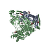
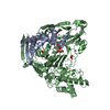
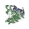
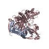
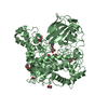
Entry Database : PDB / ID : 2wydTitle The quorum quenching N-acyl homoserine lactone acylase PvdQ in complex with dodecanoic acid ACYL-HOMOSERINE LACTONE ACYLASE PVDQ SUBUNIT ALPHA ACYL-HOMOSERINE LACTONE ACYLASE PVDQ SUBUNIT BETA Keywords / / Function / homology Function Domain/homology Component
/ / / / / / / / / / / / / / / / / / / / / / / / / / / / / / / / / / / / / / / Biological species PSEUDOMONAS AERUGINOSA (bacteria)Method / / / Resolution : 1.901 Å Authors Bokhove, M. / Nadal Jimenez, P. / Quax, W.J. / Dijkstra, B.W. Journal : Proc.Natl.Acad.Sci.USA / Year : 2010Title : The Quorum-Quenching N-Acyl Homoserine Lactone Acylase Pvdq is an Ntn-Hydrolase with an Unusual Substrate-Binding PocketAuthors : Bokhove, M. / Nadal Jimenez, P. / Quax, W.J. / Dijkstra, B.W. History Deposition Nov 16, 2009 Deposition site / Processing site Revision 1.0 Dec 29, 2009 Provider / Type Revision 1.1 May 8, 2011 Group Revision 1.2 Jul 13, 2011 Group Revision 1.3 Nov 13, 2024 Group Data collection / Database references ... Data collection / Database references / Derived calculations / Other / Structure summary Category chem_comp_atom / chem_comp_bond ... chem_comp_atom / chem_comp_bond / database_2 / pdbx_database_status / pdbx_entry_details / pdbx_modification_feature / struct_site Item _database_2.pdbx_DOI / _database_2.pdbx_database_accession ... _database_2.pdbx_DOI / _database_2.pdbx_database_accession / _pdbx_database_status.status_code_sf / _struct_site.pdbx_auth_asym_id / _struct_site.pdbx_auth_comp_id / _struct_site.pdbx_auth_seq_id
Show all Show less
 Movie
Movie Controller

 Yorodumi
 Open data
 Basic information
 Components
 Keywords
 Function and homology information
 PSEUDOMONAS AERUGINOSA (bacteria)
 X-RAY DIFFRACTION /
  Authors
 Citation
 Structure visualization
 Downloads & links
 Other downloads
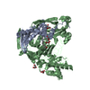


 PDBj
PDBj


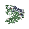
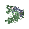
 Assembly
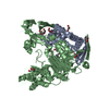

 Mass: 92.094 Da / Num. of mol.: 8 / Source method: obtained synthetically / Formula: C3H8O3
 Mass: 200.318 Da / Num. of mol.: 2 / Source method: obtained synthetically / Formula: C12H24O2
 Mass: 18.015 Da / Num. of mol.: 432 / Source method: isolated from a natural source / Formula: H2O
 Sample preparation
 / Beamline: BM16 / Wavelength: 0.979
  Processing