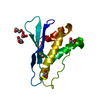
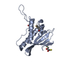
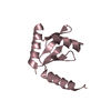
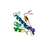
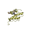
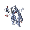
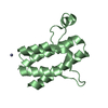
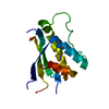
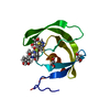
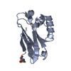
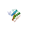
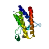
[English] 日本語
Yorodumi
- PDB-2wwe: Crystal structure of the phox homology domain of human phosphoino... -

+
Open data


ID or keywords:

Loading...

-
Basic information

Entry
Database: PDB / ID: 2wwe
TitleCrystal structure of the phox homology domain of human phosphoinositide-3-kinase-C2-gamma
ComponentsPHOSPHOINOSITIDE-3-KINASE, CLASS 2, GAMMA POLYPEPTIDE
KeywordsTRANSFERASE / PHOSPHOPROTEIN / NUCLEOTIDE-BINDING / KINASE / PIK3C2G / MEMBRANE / PX-DOMAIN / ATP-BINDING / POLYMORPHISM / CLASS 2 PI3K
Function / homology
Function and homology information


: / : / Synthesis of PIPs at the Golgi membrane / phosphatidylinositol-4-phosphate 3-kinase / phosphatidylinositol kinase activity / phosphatidylinositol biosynthetic process / phosphatidylinositol 3-kinase complex / 1-phosphatidylinositol-4-phosphate 3-kinase activity / phosphatidylinositol-3-phosphate biosynthetic process / 1-phosphatidylinositol-3-kinase activity ...: / : / Synthesis of PIPs at the Golgi membrane / phosphatidylinositol-4-phosphate 3-kinase / phosphatidylinositol kinase activity / phosphatidylinositol biosynthetic process / phosphatidylinositol 3-kinase complex / 1-phosphatidylinositol-4-phosphate 3-kinase activity / phosphatidylinositol-3-phosphate biosynthetic process / 1-phosphatidylinositol-3-kinase activity / Synthesis of PIPs at the plasma membrane / phosphatidylinositol-mediated signaling / phosphatidylinositol phosphate biosynthetic process / phosphatidylinositol binding / chemotaxis / cell migration / viral RNA genome replication / ATP binding / membrane / plasma membrane / cytoplasm / cytosol
Similarity search - Function
PI3K-C2-gamma, catalytic domain / Phox-like domain / PX Domain / PhoX homologous domain, present in p47phox and p40phox. / PX domain profile. / PX domain / Phox homology / PX domain superfamily / Phosphatidylinositol 3-kinase Ras-binding (PI3K RBD) domain / PI3-kinase family, ras-binding domain ...PI3K-C2-gamma, catalytic domain / Phox-like domain / PX Domain / PhoX homologous domain, present in p47phox and p40phox. / PX domain profile. / PX domain / Phox homology / PX domain superfamily / Phosphatidylinositol 3-kinase Ras-binding (PI3K RBD) domain / PI3-kinase family, ras-binding domain / Phosphatidylinositol 3-kinase Ras-binding (PI3K RBD) domain profile. / C2 phosphatidylinositol 3-kinase-type domain / C2 phosphatidylinositol 3-kinase (PI3K)-type domain profile. / Phosphoinositide 3-kinase, accessory (PIK) domain superfamily / Phosphoinositide 3-kinase family, accessory domain (PIK domain) / Phosphoinositide 3-kinase family, accessory domain (PIK domain) / Phosphoinositide 3-kinase, accessory (PIK) domain / Phosphatidylinositol kinase / PIK helical domain profile. / C2 domain / Protein kinase C conserved region 2 (CalB) / C2 domain / C2 domain profile. / Phosphatidylinositol 3- and 4-kinases signature 1. / Phosphatidylinositol 3/4-kinase, conserved site / Phosphatidylinositol 3- and 4-kinases signature 2. / Phosphatidylinositol 3-/4-kinase, catalytic domain superfamily / Phosphoinositide 3-kinase, catalytic domain / Phosphatidylinositol 3- and 4-kinase / Phosphatidylinositol 3- and 4-kinases catalytic domain profile. / Phosphatidylinositol 3-/4-kinase, catalytic domain / C2 domain superfamily / Armadillo-type fold / Ubiquitin-like domain superfamily / Protein kinase-like domain superfamily / 2-Layer Sandwich / Alpha Beta
Similarity search - Domain/homology
Phosphatidylinositol 4-phosphate 3-kinase C2 domain-containing subunit gamma / Phosphatidylinositol 4-phosphate 3-kinase C2 domain-containing subunit gamma
Similarity search - Component
Biological speciesHOMO SAPIENS (human)
MethodX-RAY DIFFRACTION / SYNCHROTRON / MOLECULAR REPLACEMENT / Resolution: 1.25 Å
AuthorsRoos, A.K. / Tresaugues, L. / Arrowsmith, C.H. / Berglund, H. / Bountra, C. / Collins, R. / Edwards, A.M. / Flodin, S. / Flores, A. / Graslund, S. ...Roos, A.K. / Tresaugues, L. / Arrowsmith, C.H. / Berglund, H. / Bountra, C. / Collins, R. / Edwards, A.M. / Flodin, S. / Flores, A. / Graslund, S. / Hammarstrom, M. / Johansson, A. / Johansson, I. / Kallas, A. / Karlberg, T. / Kotyenova, T. / Kotzch, A. / Kraulis, P. / Markova, N. / Moche, M. / Nielsen, T.K. / Nyman, T. / Persson, C. / Schuler, H. / Schutz, P. / Siponen, M.I. / Svensson, L. / Thorsell, A.G. / Van Der Berg, S. / Wahlberg, E. / Weigelt, J. / Welin, M. / Wisniewska, M. / Nordlund, P. / Structural Genomics Consortium (SGC)
CitationJournal: To be Published
Title: Crystal Structure of the Phox Homology Domain of Human Phosphoinositide-3-Kinase-C2-Gamma
Authors: Roos, A.K. / Tresaugues, L. / Arrowsmith, C.H. / Berglund, H. / Bountra, C. / Collins, R. / Edwards, A.M. / Flodin, S. / Flores, A. / Graslund, S. / Hammarstrom, M. / Johansson, A. / ...Authors: Roos, A.K. / Tresaugues, L. / Arrowsmith, C.H. / Berglund, H. / Bountra, C. / Collins, R. / Edwards, A.M. / Flodin, S. / Flores, A. / Graslund, S. / Hammarstrom, M. / Johansson, A. / Johansson, I. / Kallas, A. / Karlberg, T. / Kotyenova, T. / Kotzch, A. / Kraulis, P. / Markova, N. / Moche, M. / Nielsen, T.K. / Nyman, T. / Persson, C. / Schuler, H. / Schutz, P. / Siponen, M.I. / Svensson, L. / Thorsell, A.G. / Van Den Berg, S. / Wahlberg, E. / Weigelt, J. / Welin, M. / Wisniewska, M. / Nordlund, P. / Structural Genomics Consortium (SGC)
History
DepositionOct 22, 2009Deposition site: PDBE / Processing site: PDBE
Revision 1.0Nov 3, 2009Provider: repository / Type: Initial release
Revision 1.1Apr 29, 2015Group: Derived calculations / Refinement description / Version format compliance
Revision 1.2May 24, 2017Group: Database references
Revision 1.3Dec 20, 2023Group: Data collection / Database references ...Data collection / Database references / Other / Refinement description
Category: chem_comp_atom / chem_comp_bond ...chem_comp_atom / chem_comp_bond / database_2 / pdbx_database_status / pdbx_initial_refinement_model
Item: _database_2.pdbx_DOI / _database_2.pdbx_database_accession / _pdbx_database_status.status_code_sf
Remark 650 HELIX DETERMINATION METHOD: AUTHOR PROVIDED.
Remark 700 SHEET DETERMINATION METHOD: AUTHOR PROVIDED.

-
Structure visualization

Structure viewerMolecule:
MolmilJmol/JSmol

Downloads & links

-
Assembly

Deposited unit
A: PHOSPHOINOSITIDE-3-KINASE, CLASS 2, GAMMA POLYPEPTIDE


Theoretical massNumber of molelcules
Total (without water)14,9781
Polymers14,9781
Non-polymers00
Water3,153175
1


  • Idetical with deposited unit
  • defined by author&software
TypeNameSymmetry operationNumber
identity operation1_555x,y,z1
MethodPISA
Unit cell
Length a, b, c (Å)53.420, 53.420, 75.130
Angle α, β, γ (deg.)90.00, 90.00, 120.00
Int Tables number154
Space group name H-MP3221

-
Components

#1: Protein PHOSPHOINOSITIDE-3-KINASE, CLASS 2, GAMMA POLYPEPTIDE


Mass: 14977.646 Da / Num. of mol.: 1 / Fragment: PHOX HOMOLOGY DOMAIN, RESIDUES 1203-1306
Source method: isolated from a genetically manipulated source
Source: (gene. exp.) HOMO SAPIENS (human) / Plasmid: PNIC-BSA4 / Production host: ESCHERICHIA COLI (E. coli) / Strain (production host): BL21(DE3) / Variant (production host): R3 PRARE / References: UniProt: A1L3U0, UniProt: O75747*PLUS
#2: Water ChemComp-HOH / water


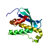
Mass: 18.015 Da / Num. of mol.: 175 / Source method: isolated from a natural source / Formula: H2O

-
Experimental details

-
Experiment

ExperimentMethod: X-RAY DIFFRACTION / Number of used crystals: 1

-
Sample preparation

CrystalDensity Matthews: 2.06 Å3/Da / Density % sol: 40.4 % / Description: NONE
Crystal growpH: 5.5
Details: 0.1M TRI-SODIUM CITRATE DIHYDRATE PH 5.5, 20% PEG3000

-
Data collection

DiffractionMean temperature: 100 K
Diffraction sourceSource: SYNCHROTRON / Site: Diamond / Beamline: I04 / Wavelength: 0.9789
DetectorType: MARRESEARCH / Detector: CCD / Date: Sep 24, 2009
RadiationProtocol: SINGLE WAVELENGTH / Monochromatic (M) / Laue (L): M / Scattering type: x-ray
Radiation wavelengthWavelength: 0.9789 Å / Relative weight: 1
ReflectionResolution: 1.25→29.16 Å / Num. obs: 34900 / % possible obs: 99.9 % / Observed criterion σ(I): 4.5 / Redundancy: 8.6 % / Biso Wilson estimate: 10 Å2 / Rmerge(I) obs: 0.07 / Net I/σ(I): 16.8
Reflection shellResolution: 1.25→1.32 Å / Redundancy: 7.1 % / Rmerge(I) obs: 0.41 / Mean I/σ(I) obs: 4.5 / % possible all: 99.5

-
Processing

Software
NameVersionClassification
REFMAC5.5.0102refinement
MOSFLMdata reduction
SCALAdata scaling
PHASERphasing
RefinementMethod to determine structure: MOLECULAR REPLACEMENT
Starting model: PDB ENTRIES 2AR5, 2IWL,1O7K, 2V6V
Resolution: 1.25→26.71 Å / Cor.coef. Fo:Fc: 0.968 / Cor.coef. Fo:Fc free: 0.959 / SU B: 0.556 / SU ML: 0.025 / Cross valid method: THROUGHOUT / ESU R: 0.042 / ESU R Free: 0.044 / Stereochemistry target values: MAXIMUM LIKELIHOOD
Details: HYDROGENS HAVE BEEN ADDED IN THE RIDING POSITIONS. U VALUES REFINED INDIVIDUALLY.
RfactorNum. reflection% reflectionSelection details
Rfree0.18398 1772 5.1 %RANDOM
Rwork0.16502 ---
obs0.16596 33091 99.83 %-
Solvent computationIon probe radii: 0.8 Å / Shrinkage radii: 0.8 Å / VDW probe radii: 1.4 Å / Solvent model: MASK
Displacement parametersBiso mean: 9.5 Å2
Baniso -1Baniso -2Baniso -3
1-0.02 Å20.01 Å20 Å2
2--0.02 Å20 Å2
3----0.03 Å2
Refinement stepCycle: LAST / Resolution: 1.25→26.71 Å
ProteinNucleic acidLigandSolventTotal
Num. atoms920 0 0 175 1095
Refine LS restraints
Refine-IDTypeDev idealDev ideal targetNumber
X-RAY DIFFRACTIONr_bond_refined_d0.0120.0211002
X-RAY DIFFRACTIONr_bond_other_d0.0010.02673
X-RAY DIFFRACTIONr_angle_refined_deg1.2821.9141367
X-RAY DIFFRACTIONr_angle_other_deg1.63131645
X-RAY DIFFRACTIONr_dihedral_angle_1_deg5.1485126
X-RAY DIFFRACTIONr_dihedral_angle_2_deg41.92624.28656
X-RAY DIFFRACTIONr_dihedral_angle_3_deg10.45515174
X-RAY DIFFRACTIONr_dihedral_angle_4_deg13.613155
X-RAY DIFFRACTIONr_chiral_restr0.0840.2146
X-RAY DIFFRACTIONr_gen_planes_refined0.0060.021133
X-RAY DIFFRACTIONr_gen_planes_other0.0010.02222
X-RAY DIFFRACTIONr_nbd_refined
X-RAY DIFFRACTIONr_nbd_other
X-RAY DIFFRACTIONr_nbtor_refined
X-RAY DIFFRACTIONr_nbtor_other
X-RAY DIFFRACTIONr_xyhbond_nbd_refined
X-RAY DIFFRACTIONr_xyhbond_nbd_other
X-RAY DIFFRACTIONr_metal_ion_refined
X-RAY DIFFRACTIONr_metal_ion_other
X-RAY DIFFRACTIONr_symmetry_vdw_refined
X-RAY DIFFRACTIONr_symmetry_vdw_other
X-RAY DIFFRACTIONr_symmetry_hbond_refined
X-RAY DIFFRACTIONr_symmetry_hbond_other
X-RAY DIFFRACTIONr_symmetry_metal_ion_refined
X-RAY DIFFRACTIONr_symmetry_metal_ion_other
X-RAY DIFFRACTIONr_mcbond_it0.8311.5586
X-RAY DIFFRACTIONr_mcbond_other0.2051.5229
X-RAY DIFFRACTIONr_mcangle_it1.5262958
X-RAY DIFFRACTIONr_mcangle_other
X-RAY DIFFRACTIONr_scbond_it2.1653416
X-RAY DIFFRACTIONr_scbond_other
X-RAY DIFFRACTIONr_scangle_it3.5144.5401
X-RAY DIFFRACTIONr_scangle_other
X-RAY DIFFRACTIONr_long_range_B_refined
X-RAY DIFFRACTIONr_long_range_B_other
X-RAY DIFFRACTIONr_rigid_bond_restr
X-RAY DIFFRACTIONr_sphericity_free
X-RAY DIFFRACTIONr_sphericity_bonded
LS refinement shellResolution: 1.25→1.282 Å / Total num. of bins used: 20
RfactorNum. reflection% reflection
Rfree0.252 131 -
Rwork0.255 2385 -
obs--98.78 %

+
About Yorodumi

-
News

-
Feb 9, 2022. New format data for meta-information of EMDB entries

New format data for meta-information of EMDB entries

  • Version 3 of the EMDB header file is now the official format.
  • The previous official version 1.9 will be removed from the archive.

Related info.:EMDB header

External links:wwPDB to switch to version 3 of the EMDB data model

-
Aug 12, 2020. Covid-19 info

Covid-19 info

URL: https://pdbjlvh1.pdbj.org/emnavi/covid19.php

New page: Covid-19 featured information page in EM Navigator.

Related info.:Covid-19 info / Mar 5, 2020. Novel coronavirus structure data

+
Mar 5, 2020. Novel coronavirus structure data

Novel coronavirus structure data

Related info.:Yorodumi Speices / Aug 12, 2020. Covid-19 info

External links:COVID-19 featured content - PDBj / Molecule of the Month (242):Coronavirus Proteases

+
Jan 31, 2019. EMDB accession codes are about to change! (news from PDBe EMDB page)

EMDB accession codes are about to change! (news from PDBe EMDB page)

  • The allocation of 4 digits for EMDB accession codes will soon come to an end. Whilst these codes will remain in use, new EMDB accession codes will include an additional digit and will expand incrementally as the available range of codes is exhausted. The current 4-digit format prefixed with “EMD-” (i.e. EMD-XXXX) will advance to a 5-digit format (i.e. EMD-XXXXX), and so on. It is currently estimated that the 4-digit codes will be depleted around Spring 2019, at which point the 5-digit format will come into force.
  • The EM Navigator/Yorodumi systems omit the EMD- prefix.

Related info.:Q: What is EMD? / ID/Accession-code notation in Yorodumi/EM Navigator

External links:EMDB Accession Codes are Changing Soon! / Contact to PDBj

+
Jul 12, 2017. Major update of PDB

Major update of PDB

  • wwPDB released updated PDB data conforming to the new PDBx/mmCIF dictionary.
  • This is a major update changing the version number from 4 to 5, and with Remediation, in which all the entries are updated.
  • In this update, many items about electron microscopy experimental information are reorganized (e.g. em_software).
  • Now, EM Navigator and Yorodumi are based on the updated data.

External links:wwPDB Remediation / Enriched Model Files Conforming to OneDep Data Standards Now Available in the PDB FTP Archive

-
Yorodumi

Thousand views of thousand structures

  • Yorodumi is a browser for structure data from EMDB, PDB, SASBDB, etc.
  • This page is also the successor to EM Navigator detail page, and also detail information page/front-end page for Omokage search.
  • The word "yorodu" (or yorozu) is an old Japanese word meaning "ten thousand". "mi" (miru) is to see.

Related info.:EMDB / PDB / SASBDB / Comparison of 3 databanks / Yorodumi Search / Aug 31, 2016. New EM Navigator & Yorodumi / Yorodumi Papers / Jmol/JSmol / Function and homology information / Changes in new EM Navigator and Yorodumi

Read more