| Entry | Database: PDB / ID: 2wvn
|
|---|
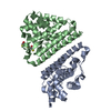
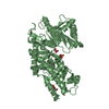
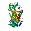
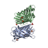
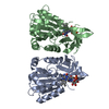
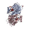
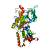
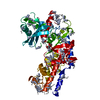
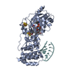
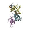
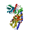
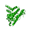
| Title | Structure of the HET-s N-terminal domain |
|---|
 Components Components | SMALL S PROTEIN |
|---|
 Keywords Keywords | PRION-BINDING PROTEIN / PRION REGULATORY DOMAIN / HETEROKARYON INCOMPATIBILITY |
|---|
| Function / homology |  Function and homology information Function and homology information
Prion-inhibition and propagation, HeLo domain / Het-s prion-forming domain / Prion-inhibition and propagation, HeLo domain / HeLo domain superfamily / Het-s 218-289 / Prion-inhibition and propagation / Four Helix Bundle (Hemerythrin (Met), subunit A) / Up-down Bundle / Mainly AlphaSimilarity search - Domain/homology |
|---|
| Biological species |  PODOSPORA ANSERINA (fungus) PODOSPORA ANSERINA (fungus) |
|---|
| Method |  X-RAY DIFFRACTION / SYNCHROTRON / MIRAS / Resolution: 2.62 Å X-RAY DIFFRACTION / SYNCHROTRON / MIRAS / Resolution: 2.62 Å |
|---|
 Authors Authors | Greenwald, J. / Buhtz, C. / Ritter, C. / Kwiatkowski, W. / Choe, S. / Saupe, S.J. / Riek, R. |
|---|
 Citation Citation | Journal: Mol.Cell / Year: 2010
Title: The Mechanism of Prion Inhibition by Het-S.
Authors: Greenwald, J. / Buhtz, C. / Ritter, C. / Kwiatkowski, W. / Choe, S. / Maddelein, M.L. / Ness, F. / Cescau, S. / Soragni, A. / Leitz, D. / Saupe, S.J. / Riek, R. |
|---|
| History | | Deposition | Oct 19, 2009 | Deposition site: PDBE / Processing site: PDBE |
|---|
| Revision 1.0 | Jul 28, 2010 | Provider: repository / Type: Initial release |
|---|
| Revision 1.1 | May 8, 2011 | Group: Version format compliance |
|---|
| Revision 1.2 | Jul 13, 2011 | Group: Version format compliance |
|---|
| Revision 1.3 | May 8, 2019 | Group: Data collection / Experimental preparation / Other
Category: exptl_crystal_grow / pdbx_database_proc / pdbx_database_status
Item: _exptl_crystal_grow.temp / _pdbx_database_status.recvd_author_approval |
|---|
| Revision 1.4 | May 8, 2024 | Group: Data collection / Database references / Other
Category: chem_comp_atom / chem_comp_bond ...chem_comp_atom / chem_comp_bond / database_2 / pdbx_database_status
Item: _database_2.pdbx_DOI / _database_2.pdbx_database_accession / _pdbx_database_status.status_code_sf |
|---|
|
|---|
 Movie
Movie Controller


 Open data
 Basic information
 Structure visualization
 Downloads & links
 Other downloads
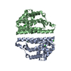
 PDBj
PDBj

 Assembly
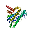

 Sample preparation
 / Beamline: X06SA / Wavelength: 1
  Processing