 Movie
Movie Controller

[English] 日本語
 Yorodumi
- PDB-2wt0: Galectin domain of porcine adenovirus type 4 NADC-1 isolate fibre... -
+ Open data

- Basic information
| Entry | Database: PDB / ID: 2wt0 | |||||||||
|---|---|---|---|---|---|---|---|---|---|---|
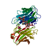
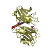
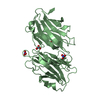
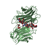
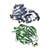
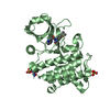
| Title | Galectin domain of porcine adenovirus type 4 NADC-1 isolate fibre complexed with N-acetyl-lactosamine | |||||||||
 Components Components | PUTATIVE FIBER PROTEIN | |||||||||
 Keywords Keywords | VIRAL PROTEIN | |||||||||
| Function / homology |  Function and homology information Function and homology informationgalactoside binding / viral capsid / carbohydrate binding / cell adhesion / symbiont entry into host cell / virion attachment to host cell Similarity search - Function | |||||||||
| Biological species |  PORCINE ADENOVIRUS 4 PORCINE ADENOVIRUS 4 | |||||||||
| Method |  X-RAY DIFFRACTION / SYNCHROTRON / MOLECULAR REPLACEMENT / Resolution: 1.91 Å X-RAY DIFFRACTION / SYNCHROTRON / MOLECULAR REPLACEMENT / Resolution: 1.91 Å | |||||||||
 Authors Authors | Guardado-Calvo, P. / Munoz, E.M. / Llamas-Saiz, A.L. / Fox, G.C. / Glasgow, J.N. / van Raaij, M.J. | |||||||||
 Citation Citation | Journal: J. Virol. / Year: 2010 Title: Crystallographic structure of porcine adenovirus type 4 fiber head and galectin domains. Authors: Guardado-Calvo, P. / Munoz, E.M. / Llamas-Saiz, A.L. / Fox, G.C. / Kahn, R. / Curiel, D.T. / Glasgow, J.N. / van Raaij, M.J. #1: Journal: Virus Res. / Year: 1995 Title: Sequence Analysis of the Fiber Genomic Region of a Porcine Adenovirus Predicts a Novel Fiber Protein. Authors: Kleiboeker, S.B. #2: Journal: Cancer Biol.Ther. / Year: 2008 Title: Characterization of Infectivity of Knob-Modified Adenoviral Vectors in Glioma. Authors: Paul, C.P.L. / Everts, M. / Glasgow, J.N. / Dent, P. / Fisher, P.B. / Ulasov, I.V. / Lesniak, M.S. / Stoff-Khalili, M.A. / Roth, J.C. / Preuss, M.A. / Dirven, C.M.F. / Lamfers, M.L.M. / ...Authors: Paul, C.P.L. / Everts, M. / Glasgow, J.N. / Dent, P. / Fisher, P.B. / Ulasov, I.V. / Lesniak, M.S. / Stoff-Khalili, M.A. / Roth, J.C. / Preuss, M.A. / Dirven, C.M.F. / Lamfers, M.L.M. / Siegal, G.P. / Zhu, Z.B. / Curiel, D.T. #3: Journal: Acta Crystallogr. Sect. F Struct. Biol. Cryst. Commun. Year: 2009 Title: Crystallization of the head and galectin-like domains of porcine adenovirus isolate NADC-1 fibre. Authors: Guardado-Calvo, P. / Llamas-Saiz, A.L. / Fox, G.C. / Glasgow, J.N. / van Raaij, M.J. | |||||||||
| History |
|
- Structure visualization
| Structure viewer | Molecule: MolmilJmol/JSmol |
|---|
- Downloads & links
-Download
| PDBx/mmCIF format | 2wt0.cif.gz | 144.1 KB | Display | PDBx/mmCIF format |
|---|---|---|---|---|
| PDB format | pdb2wt0.ent.gz | 110.4 KB | Display | PDB format |
| PDBx/mmJSON format | 2wt0.json.gz | Tree view | PDBx/mmJSON format | |
| Others |  Other downloads Other downloads |
-Validation report
| Arichive directory | https://data.pdbj.org/pub/pdb/validation_reports/wt/2wt0ftp://data.pdbj.org/pub/pdb/validation_reports/wt/2wt0 | HTTPS FTP |
|---|
-Related structure data
| Related structure data | 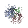 2wstC  2wsuSC  2wsvC  2wt1C  2wt2C C: citing same article ( S: Starting model for refinement |
|---|---|
| Similar structure data |


-Links
 PDBj
PDBj
- Assembly
| Deposited unit | 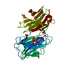
| ||||||||
|---|---|---|---|---|---|---|---|---|---|
| 1 |
| ||||||||
| Unit cell |
|
-Components
| #1: Protein | Mass: 37909.156 Da / Num. of mol.: 1 / Fragment: GALECTIN DOMAIN, RESIDUES 393-703 Source method: isolated from a genetically manipulated source Source: (gene. exp.) PORCINE ADENOVIRUS 4 / Variant: NADC-1 ISOLATE / Plasmid: PET28C / Production host:  | ||||
|---|---|---|---|---|---|
| #2: Polysaccharide | beta-D-galactopyranose-(1-4)-2-acetamido-2-deoxy-beta-D-glucopyranose Source method: isolated from a genetically manipulated source | ||||
| #3: Chemical | ChemComp-NO3 /   Mass: 62.005 Da / Num. of mol.: 4 / Source method: obtained synthetically / Formula: NO3 Mass: 62.005 Da / Num. of mol.: 4 / Source method: obtained synthetically / Formula: NO3#4: Water | ChemComp-HOH / |  Mass: 18.015 Da / Num. of mol.: 163 / Source method: isolated from a natural source / Formula: H2O Mass: 18.015 Da / Num. of mol.: 163 / Source method: isolated from a natural source / Formula: H2ONonpolymer details | BETA-D-GALACTOSE (GAL): PART OF OLIGOMER N-ACETYL-D-GLUCOSAMIN | |
-Experimental details
-Experiment
| Experiment | Method: X-RAY DIFFRACTION / Number of used crystals: 1 |
|---|
- Sample preparation
| Crystal | Density Matthews: 1.92 Å3/Da / Density % sol: 36 % / Description: NONE |
|---|---|
| Crystal grow | pH: 8 Details: 35% (W/V) PEG 3350, 500 MM SODIUM NITRATE, 5 MM DITHIOTHREITOL, 40 MM N-ACETYL-LACTOSAMINE, 10 MM TRIS-HCL 8.0 |
-Data collection
| Diffraction | Mean temperature: 100 K |
|---|---|
| Diffraction source | Source: SYNCHROTRON / Site: EMBL/DESY, HAMBURG  / Beamline: X11 / Wavelength: 0.815 / Beamline: X11 / Wavelength: 0.815 |
| Detector | Type: MAR555 FLAT PANEL / Detector: IMAGE PLATE / Date: Feb 17, 2009 / Details: BENT, VERTICALLY FOCUSSING MIRROR |
| Radiation | Monochromator: SI(111) / Protocol: SINGLE WAVELENGTH / Monochromatic (M) / Laue (L): M / Scattering type: x-ray |
| Radiation wavelength | Wavelength: 0.815 Å / Relative weight: 1 |
| Reflection | Resolution: 1.91→35 Å / Num. obs: 22025 / % possible obs: 98 % / Observed criterion σ(I): 2 / Redundancy: 2.7 % / Biso Wilson estimate: 25.423 Å2 / Rmerge(I) obs: 0.1 / Net I/σ(I): 5.9 |
| Reflection shell | Resolution: 1.91→2.01 Å / Redundancy: 2.7 % / Rmerge(I) obs: 0.31 / Mean I/σ(I) obs: 2.7 / % possible all: 98.5 |
- Processing
| Software |
| ||||||||||||||||||||||||||||||||||||||||||||||||||||||||||||||||||||||||||||||||||||||||||||||||||||||||||||||||||||||||||||||||||||||||||||||||||||||||||||||||||||||||||||||||||||||
|---|---|---|---|---|---|---|---|---|---|---|---|---|---|---|---|---|---|---|---|---|---|---|---|---|---|---|---|---|---|---|---|---|---|---|---|---|---|---|---|---|---|---|---|---|---|---|---|---|---|---|---|---|---|---|---|---|---|---|---|---|---|---|---|---|---|---|---|---|---|---|---|---|---|---|---|---|---|---|---|---|---|---|---|---|---|---|---|---|---|---|---|---|---|---|---|---|---|---|---|---|---|---|---|---|---|---|---|---|---|---|---|---|---|---|---|---|---|---|---|---|---|---|---|---|---|---|---|---|---|---|---|---|---|---|---|---|---|---|---|---|---|---|---|---|---|---|---|---|---|---|---|---|---|---|---|---|---|---|---|---|---|---|---|---|---|---|---|---|---|---|---|---|---|---|---|---|---|---|---|---|---|---|---|
| Refinement | Method to determine structure: MOLECULAR REPLACEMENT Starting model: PDB ENTRY 2WSU, CHAIN A Resolution: 1.91→35 Å / Cor.coef. Fo:Fc: 0.958 / Cor.coef. Fo:Fc free: 0.932 / SU B: 8.047 / SU ML: 0.112 / Cross valid method: THROUGHOUT / ESU R: 0.175 / ESU R Free: 0.163 / Stereochemistry target values: MAXIMUM LIKELIHOOD Details: HYDROGENS HAVE BEEN ADDED IN THE RIDING POSITIONS. U VALUES WITH TLS ADDED
| ||||||||||||||||||||||||||||||||||||||||||||||||||||||||||||||||||||||||||||||||||||||||||||||||||||||||||||||||||||||||||||||||||||||||||||||||||||||||||||||||||||||||||||||||||||||
| Solvent computation | Ion probe radii: 0.8 Å / Shrinkage radii: 0.8 Å / VDW probe radii: 1.4 Å / Solvent model: MASK | ||||||||||||||||||||||||||||||||||||||||||||||||||||||||||||||||||||||||||||||||||||||||||||||||||||||||||||||||||||||||||||||||||||||||||||||||||||||||||||||||||||||||||||||||||||||
| Displacement parameters | Biso mean: 26.591 Å2
| ||||||||||||||||||||||||||||||||||||||||||||||||||||||||||||||||||||||||||||||||||||||||||||||||||||||||||||||||||||||||||||||||||||||||||||||||||||||||||||||||||||||||||||||||||||||
| Refinement step | Cycle: LAST / Resolution: 1.91→35 Å
| ||||||||||||||||||||||||||||||||||||||||||||||||||||||||||||||||||||||||||||||||||||||||||||||||||||||||||||||||||||||||||||||||||||||||||||||||||||||||||||||||||||||||||||||||||||||
| Refine LS restraints |
|