 Movie
Movie Controller


+ Open data

- Basic information
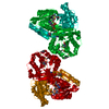
| Entry | Database: PDB / ID: 2wiu | ||||||
|---|---|---|---|---|---|---|---|
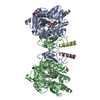
| Title | Mercury-modified bacterial persistence regulator hipBA | ||||||
 Components Components |
| ||||||
 Keywords Keywords | TRANSFERASE/TRANSCRIPTION / TRANSFERASE TRANSCRIPTION COMPLEX / SERINE KINASE / DNA-BINDING / MERCURY DERIVATIVE / REPRESSOR / TRANSCRIPTION REGULATION / SAD / TRANSFERASE-TRANSCRIPTION complex | ||||||
| Function / homology |  Function and homology information Function and homology informationdormancy process / toxin-antitoxin complex / single-species biofilm formation / regulation of growth / DNA-binding transcription repressor activity / core promoter sequence-specific DNA binding / protein-DNA complex / sequence-specific DNA binding / non-specific serine/threonine protein kinase / transcription cis-regulatory region binding ...dormancy process / toxin-antitoxin complex / single-species biofilm formation / regulation of growth / DNA-binding transcription repressor activity / core promoter sequence-specific DNA binding / protein-DNA complex / sequence-specific DNA binding / non-specific serine/threonine protein kinase / transcription cis-regulatory region binding / protein serine kinase activity / response to antibiotic / negative regulation of DNA-templated transcription / protein serine/threonine kinase activity / regulation of DNA-templated transcription / DNA-templated transcription / magnesium ion binding / protein homodimerization activity / ATP binding / cytosol Similarity search - Function | ||||||
| Biological species |  | ||||||
| Method |  X-RAY DIFFRACTION / SYNCHROTRON / SAD / Resolution: 2.35 Å X-RAY DIFFRACTION / SYNCHROTRON / SAD / Resolution: 2.35 Å | ||||||
 Authors Authors | Evdokimov, A. / Voznesensky, I. / Fennell, K. / Anderson, M. / Smith, J.F. / Fisher, D.A. | ||||||
 Citation Citation | Journal: Acta Crystallogr.,Sect.D / Year: 2009 Title: New Kinase Regulation Mechanism Found in Hipba: A Bacterial Persistence Switch. Authors: Evdokimov, A. / Voznesensky, I. / Fennell, K. / Anderson, M. / Smith, J.F. / Fisher, D.A. | ||||||
| History |
|
- Structure visualization
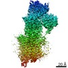
| Structure viewer | Molecule: MolmilJmol/JSmol |
|---|
- Downloads & links
-Download
| PDBx/mmCIF format | 2wiu.cif.gz | 208.2 KB | Display | PDBx/mmCIF format |
|---|---|---|---|---|
| PDB format | pdb2wiu.ent.gz | 168.2 KB | Display | PDB format |
| PDBx/mmJSON format | 2wiu.json.gz | Tree view | PDBx/mmJSON format | |
| Others |  Other downloads Other downloads |
-Validation report
| Arichive directory | https://data.pdbj.org/pub/pdb/validation_reports/wi/2wiuftp://data.pdbj.org/pub/pdb/validation_reports/wi/2wiu | HTTPS FTP |
|---|
-Related structure data
| Related structure data | |
|---|---|
| Similar structure data |


-Links
 PDBj
PDBj


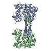
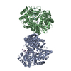
- Assembly
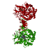
| Deposited unit | 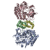
| ||||||||
|---|---|---|---|---|---|---|---|---|---|
| 1 |
| ||||||||
| Unit cell |
|
-Components
| #1: Protein | Mass: 50166.531 Da / Num. of mol.: 2 Source method: isolated from a genetically manipulated source Source: (gene. exp.) References: UniProt: P23874, non-specific serine/threonine protein kinase #2: Protein | Mass: 10024.329 Da / Num. of mol.: 2 Source method: isolated from a genetically manipulated source Source: (gene. exp.) #3: Chemical | ChemComp-HG /   Mass: 200.590 Da / Num. of mol.: 7 / Source method: obtained synthetically / Formula: Hg Mass: 200.590 Da / Num. of mol.: 7 / Source method: obtained synthetically / Formula: Hg#4: Chemical | ChemComp-CL /   Mass: 35.453 Da / Num. of mol.: 10 / Source method: obtained synthetically / Formula: Cl Mass: 35.453 Da / Num. of mol.: 10 / Source method: obtained synthetically / Formula: Cl#5: Water | ChemComp-HOH / |  Mass: 18.015 Da / Num. of mol.: 278 / Source method: isolated from a natural source / Formula: H2O Mass: 18.015 Da / Num. of mol.: 278 / Source method: isolated from a natural source / Formula: H2O |
|---|
-Experimental details
-Experiment
| Experiment | Method: X-RAY DIFFRACTION / Number of used crystals: 1 |
|---|
- Sample preparation
| Crystal | Density Matthews: 3.86 Å3/Da / Density % sol: 67.89 % Description: HG-SAD AT 1.01A USING SHARP, DENSITY MODIFIED IN SOLOMON AND RESOLVE |
|---|---|
| Crystal grow | pH: 7.5 Details: 2M AMMONIUM SULFATE, HEPES PH 7.6, 6% ETHYLENE GLYCOL 10 MG/ML PROTEIN |
-Data collection
| Diffraction | Mean temperature: 100 K |
|---|---|
| Diffraction source | Source: SYNCHROTRON / Site: NSLS  / Beamline: X25 / Wavelength: 1.01 / Beamline: X25 / Wavelength: 1.01 |
| Detector | Type: ADSC CCD / Detector: CCD / Date: Aug 6, 2008 / Details: MIRRORS |
| Radiation | Monochromator: SI / Protocol: SINGLE WAVELENGTH / Monochromatic (M) / Laue (L): M / Scattering type: x-ray |
| Radiation wavelength | Wavelength: 1.01 Å / Relative weight: 1 |
| Reflection | Resolution: 2.35→100 Å / Num. obs: 73235 / % possible obs: 99.5 % / Observed criterion σ(I): 0 / Redundancy: 8.8 % / Biso Wilson estimate: 48 Å2 / Rmerge(I) obs: 0.03 / Net I/σ(I): 23.26 |
| Reflection shell | Resolution: 2.35→2.45 Å / Redundancy: 7.8 % / Rmerge(I) obs: 0.32 / Mean I/σ(I) obs: 3.9 / % possible all: 100 |
- Processing
| Software |
| ||||||||||||||||||||||||||||||||||||||||||||||||||||||||||||||||||||||||||||||||||||||||||||||||||||||||||||||||||||||||||||||||||||||||||||||||||||||||||||||||||||||||||||||||||||||
|---|---|---|---|---|---|---|---|---|---|---|---|---|---|---|---|---|---|---|---|---|---|---|---|---|---|---|---|---|---|---|---|---|---|---|---|---|---|---|---|---|---|---|---|---|---|---|---|---|---|---|---|---|---|---|---|---|---|---|---|---|---|---|---|---|---|---|---|---|---|---|---|---|---|---|---|---|---|---|---|---|---|---|---|---|---|---|---|---|---|---|---|---|---|---|---|---|---|---|---|---|---|---|---|---|---|---|---|---|---|---|---|---|---|---|---|---|---|---|---|---|---|---|---|---|---|---|---|---|---|---|---|---|---|---|---|---|---|---|---|---|---|---|---|---|---|---|---|---|---|---|---|---|---|---|---|---|---|---|---|---|---|---|---|---|---|---|---|---|---|---|---|---|---|---|---|---|---|---|---|---|---|---|---|
| Refinement | Method to determine structure: SAD Starting model: NONE Resolution: 2.35→166.67 Å / Cor.coef. Fo:Fc: 0.944 / Cor.coef. Fo:Fc free: 0.925 / SU B: 6.764 / SU ML: 0.161 / Cross valid method: THROUGHOUT / ESU R: 0.241 / ESU R Free: 0.215 / Stereochemistry target values: MAXIMUM LIKELIHOOD Details: HYDROGENS HAVE BEEN ADDED IN THE RIDING POSITIONS. DISORDERED REGIONS WERE MOSTLY OMITTED. REGIONS IN THE VICINITY OF BOUND MERCURY IONS ARE POORLY RESOLVED DUE TO DISORDER AND PARTIAL OCCUPANCY
| ||||||||||||||||||||||||||||||||||||||||||||||||||||||||||||||||||||||||||||||||||||||||||||||||||||||||||||||||||||||||||||||||||||||||||||||||||||||||||||||||||||||||||||||||||||||
| Solvent computation | Ion probe radii: 0.8 Å / Shrinkage radii: 0.8 Å / VDW probe radii: 1.2 Å / Solvent model: MASK | ||||||||||||||||||||||||||||||||||||||||||||||||||||||||||||||||||||||||||||||||||||||||||||||||||||||||||||||||||||||||||||||||||||||||||||||||||||||||||||||||||||||||||||||||||||||
| Displacement parameters | Biso mean: 49.022 Å2
| ||||||||||||||||||||||||||||||||||||||||||||||||||||||||||||||||||||||||||||||||||||||||||||||||||||||||||||||||||||||||||||||||||||||||||||||||||||||||||||||||||||||||||||||||||||||
| Refinement step | Cycle: LAST / Resolution: 2.35→166.67 Å
| ||||||||||||||||||||||||||||||||||||||||||||||||||||||||||||||||||||||||||||||||||||||||||||||||||||||||||||||||||||||||||||||||||||||||||||||||||||||||||||||||||||||||||||||||||||||
| Refine LS restraints |
|