 Movie
Movie Controller

[English] 日本語
 Yorodumi
- PDB-2wi9: Selective oxidation of carbolide C-H bonds by engineered macrolid... -
+ Open data

- Basic information
| Entry | Database: PDB / ID: 2wi9 | ||||||
|---|---|---|---|---|---|---|---|
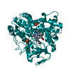
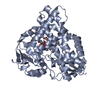
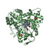
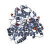
| Title | Selective oxidation of carbolide C-H bonds by engineered macrolide P450 monooxygenase | ||||||
 Components Components | CYTOCHROME P450 HYDROXYLASE PIKC | ||||||
 Keywords Keywords | OXIDOREDUCTASE / ANTIBIOTIC BIOSYNTHESIS / CYP107L1 / CYTOCHROME P450 / HEME / IRON / MACROLIDE MONOOXYGENASE / METAL-BINDING / MONOOXYGENASE / OXIDOREDUCTASE ANTIBIOTIC BIOSYNTHESIS / PIKC | ||||||
| Function / homology |  Function and homology information Function and homology informationpikromycin synthase / macrolide biosynthetic process / oxidoreductase activity, acting on paired donors, with incorporation or reduction of molecular oxygen / monooxygenase activity / iron ion binding / heme binding Similarity search - Function | ||||||
| Biological species |  STREPTOMYCES VENEZUELAE (bacteria) STREPTOMYCES VENEZUELAE (bacteria) | ||||||
| Method |  X-RAY DIFFRACTION / SYNCHROTRON / MOLECULAR REPLACEMENT / Resolution: 2 Å X-RAY DIFFRACTION / SYNCHROTRON / MOLECULAR REPLACEMENT / Resolution: 2 Å | ||||||
 Authors Authors | Li, S. / Chaulagain, M.R. / Knauff, A.R. / Podust, L.M. / Montgomery, J. / Sherman, D.H. | ||||||
 Citation Citation | Journal: Proc.Natl.Acad.Sci.USA / Year: 2009 Title: Selective Oxidation of Carbolide C-H Bonds by an Engineered Macrolide P450 Mono-Oxygenase. Authors: Li, S. / Chaulagain, M.R. / Knauff, A.R. / Podust, L.M. / Montgomery, J. / Sherman, D.H. | ||||||
| History |
|
- Structure visualization
| Structure viewer | Molecule: MolmilJmol/JSmol |
|---|
- Downloads & links
-Download
| PDBx/mmCIF format | 2wi9.cif.gz | 185.3 KB | Display | PDBx/mmCIF format |
|---|---|---|---|---|
| PDB format | pdb2wi9.ent.gz | 146.7 KB | Display | PDB format |
| PDBx/mmJSON format | 2wi9.json.gz | Tree view | PDBx/mmJSON format | |
| Others |  Other downloads Other downloads |
-Validation report
| Arichive directory | https://data.pdbj.org/pub/pdb/validation_reports/wi/2wi9ftp://data.pdbj.org/pub/pdb/validation_reports/wi/2wi9 | HTTPS FTP |
|---|
-Related structure data
| Related structure data |  2whwC  2c6hS S: Starting model for refinement C: citing same article ( |
|---|---|
| Similar structure data |


-Links
 PDBj
PDBj
- Assembly
| Deposited unit | 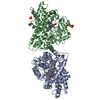
| ||||||||
|---|---|---|---|---|---|---|---|---|---|
| 1 | 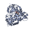
| ||||||||
| 2 | 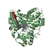
| ||||||||
| Unit cell |
| ||||||||
| Noncrystallographic symmetry (NCS) | NCS oper: (Code: given Matrix: (-0.99993, -0.01215, 0.00056), Vector: |
-Components
| #1: Protein | Mass: 48181.641 Da / Num. of mol.: 2 / Mutation: YES Source method: isolated from a genetically manipulated source Source: (gene. exp.) STREPTOMYCES VENEZUELAE (bacteria) / Plasmid: PET28A / Production host: #2: Chemical |   Mass: 616.487 Da / Num. of mol.: 2 / Source method: obtained synthetically / Formula: C34H32FeN4O4 Mass: 616.487 Da / Num. of mol.: 2 / Source method: obtained synthetically / Formula: C34H32FeN4O4#3: Chemical |   Mass: 341.529 Da / Num. of mol.: 2 / Source method: obtained synthetically / Formula: C20H39NO3 Mass: 341.529 Da / Num. of mol.: 2 / Source method: obtained synthetically / Formula: C20H39NO3#4: Chemical |   Mass: 96.063 Da / Num. of mol.: 3 / Source method: obtained synthetically / Formula: SO4 Mass: 96.063 Da / Num. of mol.: 3 / Source method: obtained synthetically / Formula: SO4#5: Water | ChemComp-HOH / |  Mass: 18.015 Da / Num. of mol.: 576 / Source method: isolated from a natural source / Formula: H2O Mass: 18.015 Da / Num. of mol.: 576 / Source method: isolated from a natural source / Formula: H2OCompound details | ENGINEERED | Sequence details | 20 N-TERMINAL RESIDUES INCLUDING 6XHIS TAG ARE ENGINEERED | |
|---|
-Experimental details
-Experiment
| Experiment | Method: X-RAY DIFFRACTION / Number of used crystals: 1 |
|---|
- Sample preparation
| Crystal | Density Matthews: 2.74 Å3/Da / Density % sol: 55.1 % / Description: NONE |
|---|---|
| Crystal grow | pH: 7.5 / Details: 15% PEG 4000, 0.1 M TRIS-HCL, PH 7.5; 200 MM MGCL2 |
-Data collection
| Diffraction | Mean temperature: 110 K |
|---|---|
| Diffraction source | Source: SYNCHROTRON / Site: ALS  / Beamline: 8.3.1 / Wavelength: 1.11587 / Beamline: 8.3.1 / Wavelength: 1.11587 |
| Detector | Type: ADSC CCD / Detector: CCD / Date: Jul 2, 2007 / Details: MIRRORS |
| Radiation | Monochromator: SI (111) DOUBLE CRYSTAL / Protocol: SINGLE WAVELENGTH / Monochromatic (M) / Laue (L): M / Scattering type: x-ray |
| Radiation wavelength | Wavelength: 1.11587 Å / Relative weight: 1 |
| Reflection | Resolution: 2→50 Å / Num. obs: 68456 / % possible obs: 98.2 % / Observed criterion σ(I): 0 / Redundancy: 87.7 % / Biso Wilson estimate: 30 Å2 / Rmerge(I) obs: 0.07 / Net I/σ(I): 21.2 |
| Reflection shell | Resolution: 2→2.07 Å / Redundancy: 3.4 % / Rmerge(I) obs: 0.39 / Mean I/σ(I) obs: 2.7 / % possible all: 87.7 |
- Processing
| Software |
| ||||||||||||||||||||||||||||||||||||||||||||||||||||||||||||||||||||||||||||||||||||||||||||||||||||||||||||||||||||||||||||||||||||||||||||||||||||||||||||||||||||||||||||||||||||||
|---|---|---|---|---|---|---|---|---|---|---|---|---|---|---|---|---|---|---|---|---|---|---|---|---|---|---|---|---|---|---|---|---|---|---|---|---|---|---|---|---|---|---|---|---|---|---|---|---|---|---|---|---|---|---|---|---|---|---|---|---|---|---|---|---|---|---|---|---|---|---|---|---|---|---|---|---|---|---|---|---|---|---|---|---|---|---|---|---|---|---|---|---|---|---|---|---|---|---|---|---|---|---|---|---|---|---|---|---|---|---|---|---|---|---|---|---|---|---|---|---|---|---|---|---|---|---|---|---|---|---|---|---|---|---|---|---|---|---|---|---|---|---|---|---|---|---|---|---|---|---|---|---|---|---|---|---|---|---|---|---|---|---|---|---|---|---|---|---|---|---|---|---|---|---|---|---|---|---|---|---|---|---|---|
| Refinement | Method to determine structure: MOLECULAR REPLACEMENT Starting model: PDB ENTRY 2C6H Resolution: 2→88.74 Å / Cor.coef. Fo:Fc: 0.956 / Cor.coef. Fo:Fc free: 0.929 / SU B: 3.931 / SU ML: 0.111 / Cross valid method: THROUGHOUT / ESU R: 0.178 / ESU R Free: 0.17 / Stereochemistry target values: MAXIMUM LIKELIHOOD / Details: HYDROGENS HAVE BEEN ADDED IN THE RIDING POSITIONS.
| ||||||||||||||||||||||||||||||||||||||||||||||||||||||||||||||||||||||||||||||||||||||||||||||||||||||||||||||||||||||||||||||||||||||||||||||||||||||||||||||||||||||||||||||||||||||
| Solvent computation | Ion probe radii: 0.8 Å / Shrinkage radii: 0.8 Å / VDW probe radii: 1.4 Å / Solvent model: MASK | ||||||||||||||||||||||||||||||||||||||||||||||||||||||||||||||||||||||||||||||||||||||||||||||||||||||||||||||||||||||||||||||||||||||||||||||||||||||||||||||||||||||||||||||||||||||
| Displacement parameters | Biso mean: 31.24 Å2
| ||||||||||||||||||||||||||||||||||||||||||||||||||||||||||||||||||||||||||||||||||||||||||||||||||||||||||||||||||||||||||||||||||||||||||||||||||||||||||||||||||||||||||||||||||||||
| Refinement step | Cycle: LAST / Resolution: 2→88.74 Å
| ||||||||||||||||||||||||||||||||||||||||||||||||||||||||||||||||||||||||||||||||||||||||||||||||||||||||||||||||||||||||||||||||||||||||||||||||||||||||||||||||||||||||||||||||||||||
| Refine LS restraints |
|