 Movie
Movie Controller


+ Open data

- Basic information
| Entry | Database: PDB / ID: 2ca0 | ||||||
|---|---|---|---|---|---|---|---|
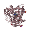
| Title | Crystal structure of YC-17-bound cytochrome P450 PikC (CYP107L1) | ||||||
 Components Components | CYTOCHROME P450 MONOOXYGENASE | ||||||
 Keywords Keywords | OXIDOREDUCTASE / CYTOCHROME P450 / PIKC / YC-17 / MACROLIDE MONOOXYGENASE / ANTIBIOTIC BIOSYNTHESIS / HEME / IRON / METAL-BINDING | ||||||
| Function / homology |  Function and homology information Function and homology informationpikromycin synthase / macrolide biosynthetic process / oxidoreductase activity, acting on paired donors, with incorporation or reduction of molecular oxygen / monooxygenase activity / iron ion binding / heme binding Similarity search - Function | ||||||
| Biological species |  STREPTOMYCES VENEZUELAE (bacteria) STREPTOMYCES VENEZUELAE (bacteria) | ||||||
| Method |  X-RAY DIFFRACTION / SYNCHROTRON / MOLECULAR REPLACEMENT / Resolution: 2.85 Å X-RAY DIFFRACTION / SYNCHROTRON / MOLECULAR REPLACEMENT / Resolution: 2.85 Å | ||||||
 Authors Authors | Yermalitskaya, L.I. / Kim, Y. / Sherman, D.H. / Waterman, M.R. / Podust, L.M. | ||||||
 Citation Citation | Journal: To be Published Title: Crystal Structure of Yc-17-Bound Cytochrome P450 Pikc (Cyp107L1) Authors: Yermalitskaya, L.I. / Kim, Y. / Sherman, D.H. / Waterman, M.R. / Podust, L.M. | ||||||
| History |
|
- Structure visualization
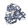
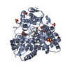
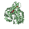
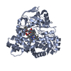
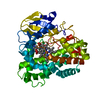
| Structure viewer | Molecule: MolmilJmol/JSmol |
|---|
- Downloads & links
-Download
| PDBx/mmCIF format | 2ca0.cif.gz | 169.1 KB | Display | PDBx/mmCIF format |
|---|---|---|---|---|
| PDB format | pdb2ca0.ent.gz | 132.3 KB | Display | PDB format |
| PDBx/mmJSON format | 2ca0.json.gz | Tree view | PDBx/mmJSON format | |
| Others |  Other downloads Other downloads |
-Validation report
| Arichive directory | https://data.pdbj.org/pub/pdb/validation_reports/ca/2ca0ftp://data.pdbj.org/pub/pdb/validation_reports/ca/2ca0 | HTTPS FTP |
|---|
-Related structure data
| Related structure data |  2bvjS S: Starting model for refinement |
|---|---|
| Similar structure data |


-Links
 PDBj
PDBj
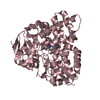
- Assembly
| Deposited unit | 
| ||||||||
|---|---|---|---|---|---|---|---|---|---|
| 1 | 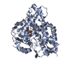
| ||||||||
| 2 | 
| ||||||||
| Unit cell |
| ||||||||
| Noncrystallographic symmetry (NCS) | NCS oper: (Code: given Matrix: (-0.99882, 0.03381, -0.03492), Vector: |
-Components
| #1: Protein | Mass: 48268.742 Da / Num. of mol.: 2 Source method: isolated from a genetically manipulated source Source: (gene. exp.) STREPTOMYCES VENEZUELAE (bacteria) / Plasmid: PET28A / Production host: #2: Chemical |   Mass: 616.487 Da / Num. of mol.: 2 / Source method: obtained synthetically / Formula: C34H32FeN4O4 Mass: 616.487 Da / Num. of mol.: 2 / Source method: obtained synthetically / Formula: C34H32FeN4O4#3: Chemical | 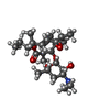  Mass: 453.612 Da / Num. of mol.: 2 / Source method: obtained synthetically / Formula: C25H43NO6 Mass: 453.612 Da / Num. of mol.: 2 / Source method: obtained synthetically / Formula: C25H43NO6#4: Water | ChemComp-HOH / |  Mass: 18.015 Da / Num. of mol.: 63 / Source method: isolated from a natural source / Formula: H2O Mass: 18.015 Da / Num. of mol.: 63 / Source method: isolated from a natural source / Formula: H2OSequence details | FIRST 20 RESIDUES INCLUDING 6X HIS-TAG AND THROMBIN CLEAVAGE SITE ARE FROM THE CLONING VECTOR PET28A | |
|---|
-Experimental details
-Experiment
| Experiment | Method: X-RAY DIFFRACTION / Number of used crystals: 1 |
|---|
- Sample preparation
| Crystal | Density Matthews: 2.21 Å3/Da / Density % sol: 43.79 % |
|---|---|
| Crystal grow | pH: 6.5 Details: 10% PEG MME 5000,0.1 M MES 6.5, 0.1 M AMMONIUM SULFATE, 0.5 MM DTT 1 MM YC-17, pH 6.50 |
-Data collection
| Diffraction | Mean temperature: 100 K |
|---|---|
| Diffraction source | Source: SYNCHROTRON / Site: APS  / Beamline: 22-BM / Wavelength: 0.97952 / Beamline: 22-BM / Wavelength: 0.97952 |
| Detector | Type: MARRESEARCH / Detector: CCD / Date: Oct 28, 2005 Details: SAGITTAL FOCUSING CRYSTAL AND VERTICAL FOCUSING DOUBLE MIRROR |
| Radiation | Monochromator: SI (111) DOUBLE CRYSTAL / Protocol: SINGLE WAVELENGTH / Monochromatic (M) / Laue (L): M / Scattering type: x-ray |
| Radiation wavelength | Wavelength: 0.97952 Å / Relative weight: 1 |
| Reflection | Resolution: 2.85→50 Å / Num. obs: 17855 / % possible obs: 93.4 % / Observed criterion σ(I): 0 / Redundancy: 2.5 % / Biso Wilson estimate: 75.2 Å2 / Rmerge(I) obs: 0.1 / Net I/σ(I): 14.1 |
| Reflection shell | Resolution: 2.85→2.95 Å / Redundancy: 1.7 % / Rmerge(I) obs: 0.16 / Mean I/σ(I) obs: 3.8 / % possible all: 75.8 |
- Processing
| Software |
| ||||||||||||||||||||||||||||||||||||||||||||||||||||||||||||||||||||||||||||||||
|---|---|---|---|---|---|---|---|---|---|---|---|---|---|---|---|---|---|---|---|---|---|---|---|---|---|---|---|---|---|---|---|---|---|---|---|---|---|---|---|---|---|---|---|---|---|---|---|---|---|---|---|---|---|---|---|---|---|---|---|---|---|---|---|---|---|---|---|---|---|---|---|---|---|---|---|---|---|---|---|---|---|
| Refinement | Method to determine structure: MOLECULAR REPLACEMENT Starting model: PDB ENTRY 2BVJ Resolution: 2.85→32.41 Å / Rfactor Rfree error: 0.008 / Data cutoff high absF: 124191.07 / Isotropic thermal model: RESTRAINED / Cross valid method: THROUGHOUT / σ(F): 0 / Stereochemistry target values: MAXIMUM LIKELIHOOD / Details: DISORDERED REGIONS WERE OMITTED FROM THE STRUCTURE
| ||||||||||||||||||||||||||||||||||||||||||||||||||||||||||||||||||||||||||||||||
| Solvent computation | Solvent model: FLAT MODEL / Bsol: 46.1387 Å2 / ksol: 0.333374 e/Å3 | ||||||||||||||||||||||||||||||||||||||||||||||||||||||||||||||||||||||||||||||||
| Displacement parameters | Biso mean: 23.1 Å2
| ||||||||||||||||||||||||||||||||||||||||||||||||||||||||||||||||||||||||||||||||
| Refine analyze |
| ||||||||||||||||||||||||||||||||||||||||||||||||||||||||||||||||||||||||||||||||
| Refinement step | Cycle: LAST / Resolution: 2.85→32.41 Å
| ||||||||||||||||||||||||||||||||||||||||||||||||||||||||||||||||||||||||||||||||
| Refine LS restraints |
| ||||||||||||||||||||||||||||||||||||||||||||||||||||||||||||||||||||||||||||||||
| LS refinement shell | Resolution: 2.85→3.03 Å / Rfactor Rfree error: 0.027 / Total num. of bins used: 6
| ||||||||||||||||||||||||||||||||||||||||||||||||||||||||||||||||||||||||||||||||
| Xplor file |
|
