 Movie
Movie Controller

[English] 日本語
 Yorodumi
- PDB-2waw: crystal structure of Mycobacterium tuberculosis rv0371c homolog f... -
+ Open data

- Basic information
| Entry | Database: PDB / ID: 2waw | ||||||
|---|---|---|---|---|---|---|---|
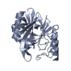
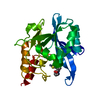
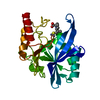
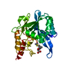
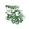
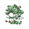
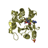
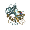
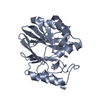
| Title | crystal structure of Mycobacterium tuberculosis rv0371c homolog from mycobacterium sp. strain JC1 | ||||||
 Components Components | MOBA RELATE PROTEIN | ||||||
 Keywords Keywords | UNKNOWN FUNCTION | ||||||
| Function / homology |  Function and homology information Function and homology information | ||||||
| Biological species |  MYCOBACTERIUM SP. JC-1 (bacteria) MYCOBACTERIUM SP. JC-1 (bacteria) | ||||||
| Method |  X-RAY DIFFRACTION / SYNCHROTRON / MOLECULAR REPLACEMENT / Resolution: 1.6 Å X-RAY DIFFRACTION / SYNCHROTRON / MOLECULAR REPLACEMENT / Resolution: 1.6 Å | ||||||
 Authors Authors | Cho, H.J. / Kang, B.S. | ||||||
 Citation Citation | Journal: To be Published Title: Crystal Structure of Mycobacterium Tuberculosis Rv0371C Homolog from Mycobacterium Sp. Strain Jc1 Authors: Cho, H.J. / Kang, B.S. | ||||||
| History |
|
- Structure visualization
| Structure viewer | Molecule: MolmilJmol/JSmol |
|---|
- Downloads & links
-Download
| PDBx/mmCIF format | 2waw.cif.gz | 56.1 KB | Display | PDBx/mmCIF format |
|---|---|---|---|---|
| PDB format | pdb2waw.ent.gz | 39.6 KB | Display | PDB format |
| PDBx/mmJSON format | 2waw.json.gz | Tree view | PDBx/mmJSON format | |
| Others |  Other downloads Other downloads |
-Validation report
| Summary document | 2waw_validation.pdf.gz | 436.8 KB | Display | wwPDB validaton report |
|---|---|---|---|---|
| Full document | 2waw_full_validation.pdf.gz | 437.8 KB | Display | |
| Data in XML | 2waw_validation.xml.gz | 12.1 KB | Display | |
| Data in CIF | 2waw_validation.cif.gz | 17.3 KB | Display | |
| Arichive directory | https://data.pdbj.org/pub/pdb/validation_reports/wa/2wawftp://data.pdbj.org/pub/pdb/validation_reports/wa/2waw | HTTPS FTP |
-Related structure data
| Similar structure data |
|---|

-Links
 PDBj
PDBj

- Assembly
| Deposited unit | 
| ||||||||
|---|---|---|---|---|---|---|---|---|---|
| 1 |
| ||||||||
| Unit cell |
|
-Components
| #1: Protein | Mass: 21128.186 Da / Num. of mol.: 1 / Source method: isolated from a natural source / Source: (natural) MYCOBACTERIUM SP. JC-1 (bacteria) / References: UniProt: D5MNX3*PLUS | ||||
|---|---|---|---|---|---|
| #2: Chemical | ChemComp-PGE /   Mass: 150.173 Da / Num. of mol.: 1 / Source method: obtained synthetically / Formula: C6H14O4 Mass: 150.173 Da / Num. of mol.: 1 / Source method: obtained synthetically / Formula: C6H14O4 | ||||
| #3: Chemical |   Mass: 35.453 Da / Num. of mol.: 2 / Source method: obtained synthetically / Formula: Cl Mass: 35.453 Da / Num. of mol.: 2 / Source method: obtained synthetically / Formula: Cl#4: Water | ChemComp-HOH / |  Mass: 18.015 Da / Num. of mol.: 203 / Source method: isolated from a natural source / Formula: H2O Mass: 18.015 Da / Num. of mol.: 203 / Source method: isolated from a natural source / Formula: H2OHas protein modification | Y | |
-Experimental details
-Experiment
| Experiment | Method: X-RAY DIFFRACTION / Number of used crystals: 1 |
|---|
- Sample preparation
| Crystal | Density Matthews: 2.2 Å3/Da / Density % sol: 44.21 % / Description: NONE |
|---|---|
| Crystal grow | pH: 7.5 Details: 28% PEG 400, 0.1M HEPES PH 7.5, 0.2M CALCIUM CHLORIDE |
-Data collection
| Diffraction | Mean temperature: 294 K |
|---|---|
| Diffraction source | Source: SYNCHROTRON / Site: PAL/PLS  / Beamline: 6C1 / Wavelength: 1.23985 / Beamline: 6C1 / Wavelength: 1.23985 |
| Detector | Type: ADSC CCD / Detector: CCD / Date: Oct 12, 2008 / Details: MIRRORS |
| Radiation | Protocol: SINGLE WAVELENGTH / Monochromatic (M) / Laue (L): M / Scattering type: x-ray |
| Radiation wavelength | Wavelength: 1.23985 Å / Relative weight: 1 |
| Reflection | Resolution: 1.58→50 Å / Num. obs: 23465 / % possible obs: 94.1 % / Redundancy: 8.9 % / Rmerge(I) obs: 0.07 / Net I/σ(I): 42.8 |
| Reflection shell | Resolution: 1.58→1.64 Å / Redundancy: 4.3 % / Rmerge(I) obs: 0.24 / Mean I/σ(I) obs: 2.8 / % possible all: 72.4 |
- Processing
| Software |
| ||||||||||||||||||||||||||||||||||||||||||||||||||||||||||||||||||||||||||||||||||||||||||||||||||||||||||||||||||||||||||||||||||||||||||||||||||||||||||||||||||||||||||||||||||||||
|---|---|---|---|---|---|---|---|---|---|---|---|---|---|---|---|---|---|---|---|---|---|---|---|---|---|---|---|---|---|---|---|---|---|---|---|---|---|---|---|---|---|---|---|---|---|---|---|---|---|---|---|---|---|---|---|---|---|---|---|---|---|---|---|---|---|---|---|---|---|---|---|---|---|---|---|---|---|---|---|---|---|---|---|---|---|---|---|---|---|---|---|---|---|---|---|---|---|---|---|---|---|---|---|---|---|---|---|---|---|---|---|---|---|---|---|---|---|---|---|---|---|---|---|---|---|---|---|---|---|---|---|---|---|---|---|---|---|---|---|---|---|---|---|---|---|---|---|---|---|---|---|---|---|---|---|---|---|---|---|---|---|---|---|---|---|---|---|---|---|---|---|---|---|---|---|---|---|---|---|---|---|---|---|
| Refinement | Method to determine structure: MOLECULAR REPLACEMENT Starting model: MYCOBACTERIUM TUBERCULOSIS RV0371C Resolution: 1.6→24.5 Å / Cor.coef. Fo:Fc: 0.965 / Cor.coef. Fo:Fc free: 0.96 / SU B: 3.247 / SU ML: 0.058 / TLS residual ADP flag: LIKELY RESIDUAL / Cross valid method: THROUGHOUT / ESU R: 0.094 / ESU R Free: 0.091 / Stereochemistry target values: MAXIMUM LIKELIHOOD / Details: HYDROGENS HAVE BEEN ADDED IN THE RIDING POSITIONS.
| ||||||||||||||||||||||||||||||||||||||||||||||||||||||||||||||||||||||||||||||||||||||||||||||||||||||||||||||||||||||||||||||||||||||||||||||||||||||||||||||||||||||||||||||||||||||
| Solvent computation | Ion probe radii: 0.8 Å / Shrinkage radii: 0.8 Å / VDW probe radii: 1.2 Å / Solvent model: MASK | ||||||||||||||||||||||||||||||||||||||||||||||||||||||||||||||||||||||||||||||||||||||||||||||||||||||||||||||||||||||||||||||||||||||||||||||||||||||||||||||||||||||||||||||||||||||
| Displacement parameters | Biso mean: 23.8 Å2
| ||||||||||||||||||||||||||||||||||||||||||||||||||||||||||||||||||||||||||||||||||||||||||||||||||||||||||||||||||||||||||||||||||||||||||||||||||||||||||||||||||||||||||||||||||||||
| Refinement step | Cycle: LAST / Resolution: 1.6→24.5 Å
| ||||||||||||||||||||||||||||||||||||||||||||||||||||||||||||||||||||||||||||||||||||||||||||||||||||||||||||||||||||||||||||||||||||||||||||||||||||||||||||||||||||||||||||||||||||||
| Refine LS restraints |
|