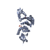
 Movie
Movie Controller


+ Open data

- Basic information
| Entry | Database: PDB / ID: 2w2g | ||||||
|---|---|---|---|---|---|---|---|
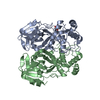
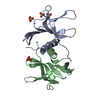
| Title | Human SARS coronavirus unique domain | ||||||
 Components Components | NON-STRUCTURAL PROTEIN 3 | ||||||
 Keywords Keywords | RNA BINDING PROTEIN / THIOL PROTEASE / RNA REPLICATION / VIRAL REPLICASE / RNA-BINDING / ZINC-FINGER / RIBOSOMAL FRAMESHIFTING / HYDROLASE / RNA-BINDING PROTEIN | ||||||
| Function / homology |  Function and homology information Function and homology informationAssembly of the SARS-CoV-1 Replication-Transcription Complex (RTC) / Maturation of replicase proteins / Transcription of SARS-CoV-1 sgRNAs / Translation of Replicase and Assembly of the Replication Transcription Complex / K48-linked deubiquitinase activity / Replication of the SARS-CoV-1 genome / K63-linked deubiquitinase activity / host cell endoplasmic reticulum / SARS-CoV-1 modulates host translation machinery / viral genome replication ...Assembly of the SARS-CoV-1 Replication-Transcription Complex (RTC) / Maturation of replicase proteins / Transcription of SARS-CoV-1 sgRNAs / Translation of Replicase and Assembly of the Replication Transcription Complex / K48-linked deubiquitinase activity / Replication of the SARS-CoV-1 genome / K63-linked deubiquitinase activity / host cell endoplasmic reticulum / SARS-CoV-1 modulates host translation machinery / viral genome replication / methyltransferase activity / SARS-CoV-1 activates/modulates innate immune responses / endonuclease activity / methylation / double membrane vesicle viral factory outer membrane / SARS coronavirus main proteinase / host cell endosome / symbiont-mediated degradation of host mRNA / mRNA guanylyltransferase / symbiont-mediated suppression of host ISG15-protein conjugation / G-quadruplex RNA binding / mRNA guanylyltransferase activity / symbiont-mediated suppression of host cytoplasmic pattern recognition receptor signaling pathway via inhibition of IRF3 activity / omega peptidase activity / symbiont-mediated perturbation of host ubiquitin-like protein modification / host cell Golgi apparatus / cysteine-type deubiquitinase activity / ubiquitinyl hydrolase 1 / Hydrolases; Acting on peptide bonds (peptidases); Cysteine endopeptidases / lyase activity / single-stranded RNA binding / viral protein processing / host cell perinuclear region of cytoplasm / symbiont-mediated suppression of host type I interferon-mediated signaling pathway / symbiont-mediated suppression of host gene expression / viral translational frameshifting / symbiont-mediated activation of host autophagy / cysteine-type endopeptidase activity / RNA-directed RNA polymerase activity / proteolysis / zinc ion binding / identical protein binding Similarity search - Function | ||||||
| Biological species |  SARS CORONAVIRUS SARS CORONAVIRUS | ||||||
| Method |  X-RAY DIFFRACTION / SYNCHROTRON / MOLECULAR REPLACEMENT / Resolution: 2.22 Å X-RAY DIFFRACTION / SYNCHROTRON / MOLECULAR REPLACEMENT / Resolution: 2.22 Å | ||||||
 Authors Authors | Tan, J. / Vonrhein, C. / Smart, O.S. / Bricogne, G. / Bollati, M. / Kusov, Y. / Hansen, G. / Mesters, J.R. / Schmidt, C.L. / Hilgenfeld, R. | ||||||
 Citation Citation | Journal: Plos Pathog. / Year: 2009 Title: The Sars-Unique Domain (Sud) of Sars Coronavirus Contains Two Macrodomains that Bind G-Quadruplexes. Authors: Tan, J. / Vonrhein, C. / Smart, O.S. / Bricogne, G. / Bollati, M. / Kusov, Y. / Hansen, G. / Mesters, J.R. / Schmidt, C.L. / Hilgenfeld, R. | ||||||
| History |
|
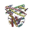
- Structure visualization
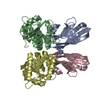
| Structure viewer | Molecule: MolmilJmol/JSmol |
|---|
- Downloads & links
-Download
| PDBx/mmCIF format | 2w2g.cif.gz | 113.6 KB | Display | PDBx/mmCIF format |
|---|---|---|---|---|
| PDB format | pdb2w2g.ent.gz | 88.3 KB | Display | PDB format |
| PDBx/mmJSON format | 2w2g.json.gz | Tree view | PDBx/mmJSON format | |
| Others |  Other downloads Other downloads |
-Validation report
| Arichive directory | https://data.pdbj.org/pub/pdb/validation_reports/w2/2w2gftp://data.pdbj.org/pub/pdb/validation_reports/w2/2w2g | HTTPS FTP |
|---|
-Related structure data
| Related structure data | 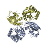 2wctC 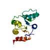 2jzeS C: citing same article ( S: Starting model for refinement |
|---|---|
| Similar structure data |


-Links
 PDBj
PDBj


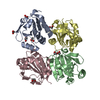
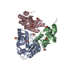
- Assembly
| Deposited unit | 
| ||||||||
|---|---|---|---|---|---|---|---|---|---|
| 1 |
| ||||||||
| Unit cell |
| ||||||||
| Noncrystallographic symmetry (NCS) | NCS oper: (Code: given Matrix: (0.82507, 0.26201, -0.50061), Vector: |
-Components
| #1: Protein | Mass: 29230.818 Da / Num. of mol.: 2 / Fragment: SARS UNIQUE DOMAIN, RESIDUES 1207-1470 / Mutation: YES Source method: isolated from a genetically manipulated source Source: (gene. exp.) SARS CORONAVIRUS / Production host:  References: UniProt: P0C6U8, Hydrolases; Acting on peptide bonds (peptidases); Cysteine endopeptidases #2: Chemical |   Mass: 96.063 Da / Num. of mol.: 2 / Source method: obtained synthetically / Formula: SO4 Mass: 96.063 Da / Num. of mol.: 2 / Source method: obtained synthetically / Formula: SO4#3: Water | ChemComp-HOH / |  Mass: 18.015 Da / Num. of mol.: 178 / Source method: isolated from a natural source / Formula: H2O Mass: 18.015 Da / Num. of mol.: 178 / Source method: isolated from a natural source / Formula: H2OCompound details | ENGINEERED | Has protein modification | Y | |
|---|
-Experimental details
-Experiment
| Experiment | Method: X-RAY DIFFRACTION / Number of used crystals: 1 |
|---|
- Sample preparation
| Crystal | Density Matthews: 2.52 Å3/Da / Density % sol: 51 % / Description: NONE |
|---|---|
| Crystal grow | pH: 6.5 Details: 20% PEGMME 5000,0.2M AMMONIUM SULFATE, 0.1M MES PH6.5 |
-Data collection
| Diffraction | Mean temperature: 100 K |
|---|---|
| Diffraction source | Source: SYNCHROTRON / Site: BESSY  / Beamline: 14.2 / Wavelength: 1.25485 / Beamline: 14.2 / Wavelength: 1.25485 |
| Detector | Type: MARMOSAIC 225 mm CCD / Detector: CCD / Date: Aug 2, 2008 / Details: MIRRORS |
| Radiation | Protocol: SINGLE WAVELENGTH / Monochromatic (M) / Laue (L): M / Scattering type: x-ray |
| Radiation wavelength | Wavelength: 1.25485 Å / Relative weight: 1 |
| Reflection | Resolution: 2.22→28.25 Å / Num. obs: 26598 / % possible obs: 92.1 % / Observed criterion σ(I): 2 / Redundancy: 6.3 % / Biso Wilson estimate: 48.34 Å2 / Rmerge(I) obs: 0.055 / Net I/σ(I): 16.9 |
| Reflection shell | Resolution: 2.22→2.34 Å / Redundancy: 2.8 % / Rmerge(I) obs: 0.373 / Mean I/σ(I) obs: 2 / % possible all: 61 |
- Processing
| Software |
| ||||||||||||||||||||
|---|---|---|---|---|---|---|---|---|---|---|---|---|---|---|---|---|---|---|---|---|---|
| Refinement | Method to determine structure: MOLECULAR REPLACEMENT Starting model: PDB ENTRY 2JZE Resolution: 2.22→27.81 Å / σ(F): 0 / Details: HYDROGENS HAVE BEEN ADDED IN THE RIDING POSITIONS.
| ||||||||||||||||||||
| Displacement parameters | Biso mean: 60.39 Å2
| ||||||||||||||||||||
| Refinement step | Cycle: LAST / Resolution: 2.22→27.81 Å
| ||||||||||||||||||||
| LS refinement shell | Resolution: 2.22→2.31 Å / Total num. of bins used: 13
|
