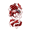
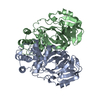
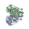
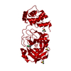
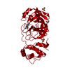
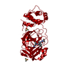
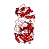
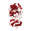
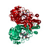
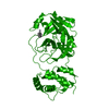
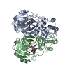
Entry Database : PDB / ID : 1q2wTitle X-Ray Crystal Structure of the SARS Coronavirus Main Protease 3C-like protease Keywords / / / Function / homology Function Domain/homology Component
/ / / / / / / / / / / / / / / / / / / / / / / / / / / / / / / / / / / / / / / / / / / / / / / / / / / / / / / / / / / / / / / / / / / / / / / / / / / / / / / / / / / / / / / / / / / / / / / / / / / / / / / / / / / / / / / / / / / / / / / / / / / / / / / / / / / / / / / / / / / / / / / / / / / / / / / / / / / / / Biological species Method / / / Resolution : 1.86 Å Authors Bonanno, J.B. / Fowler, R. / Gupta, S. / Hendle, J. / Lorimer, D. / Romero, R. / Sauder, J.M. / Wei, C.L. / Liu, E.T. / Burley, S.K. / Harris, T. Journal : New York Times / Year : 2003Title : Company Says It Mapped Part of SARS VirusAuthors : Bonanno, J.B. / Fowler, R. / Gupta, S. / Hendle, J. / Lorimer, D. / Romero, R. / Sauder, J.M. / Wei, C.L. / Liu, E.T. / Burley, S.K. / Harris, T. History Deposition Jul 26, 2003 Deposition site / Processing site Revision 1.0 Jul 29, 2003 Provider / Type Revision 1.1 Apr 29, 2008 Group Revision 1.2 Jul 13, 2011 Group Revision 1.3 Oct 11, 2017 Group / Category Revision 1.4 Nov 14, 2018 Group / Database references / Structure summaryCategory / citation_authorItem / _audit_author.name / _citation_author.nameRevision 1.5 Feb 12, 2020 Group / Source and taxonomy / Structure summaryCategory citation / entity ... citation / entity / entity_name_com / entity_src_gen / struct_ref / struct_ref_seq / struct_ref_seq_dif Item _citation.country / _citation.journal_abbrev ... _citation.country / _citation.journal_abbrev / _citation.journal_id_CSD / _citation.journal_id_ISSN / _citation.title / _citation.year / _entity.pdbx_description / _entity.pdbx_ec / _entity.pdbx_fragment / _entity_name_com.name / _entity_src_gen.gene_src_common_name / _entity_src_gen.gene_src_strain / _entity_src_gen.pdbx_beg_seq_num / _entity_src_gen.pdbx_end_seq_num / _entity_src_gen.pdbx_gene_src_gene / _entity_src_gen.pdbx_gene_src_ncbi_taxonomy_id / _entity_src_gen.pdbx_gene_src_scientific_name / _entity_src_gen.pdbx_seq_type / _struct_ref.db_code / _struct_ref.pdbx_db_accession / _struct_ref_seq.pdbx_db_accession / _struct_ref_seq_dif.details / _struct_ref_seq_dif.pdbx_seq_db_accession_code Revision 1.6 Feb 26, 2020 Group / Category Item / _citation.page_first / _citation.page_lastRevision 1.7 Feb 3, 2021 Group / Derived calculations / Structure summaryCategory / citation_author / struct_siteItem _audit_author.identifier_ORCID / _citation_author.identifier_ORCID ... _audit_author.identifier_ORCID / _citation_author.identifier_ORCID / _struct_site.pdbx_auth_asym_id / _struct_site.pdbx_auth_comp_id / _struct_site.pdbx_auth_seq_id Revision 1.8 Aug 16, 2023 Group / Database references / Refinement descriptionCategory chem_comp_atom / chem_comp_bond ... chem_comp_atom / chem_comp_bond / database_2 / pdbx_initial_refinement_model Item / _database_2.pdbx_database_accession
Show all Show less Remark 600 heterogen 15% MPD was added to the crystals as a cryoprotectant.
 Movie
Movie Controller


 Open data
 Basic information
 Components
 Keywords
 Function and homology information
 Severe acute respiratory syndrome-related coronavirus
 X-RAY DIFFRACTION /
  Authors
 Citation
 Structure visualization
 Downloads & links
 Other downloads
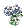


 PDBj
PDBj


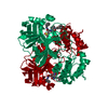
 Assembly
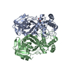


 Mass: 118.174 Da / Num. of mol.: 1 / Source method: obtained synthetically / Formula: C6H14O2 / Comment: precipitant*YM
 Mass: 18.015 Da / Num. of mol.: 456 / Source method: isolated from a natural source / Formula: H2O
 Sample preparation
 / Beamline: 31-ID / Wavelength: 0.9198 Å
 Processing