 Movie
Movie Controller

[English] 日本語
 Yorodumi
- PDB-2vko: Structure of the soluble domain of the membrane protein TM1634 fr... -
+ Open data

- Basic information
| Entry | Database: PDB / ID: 2vko | ||||||
|---|---|---|---|---|---|---|---|
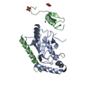
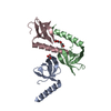
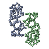
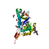
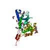
| Title | Structure of the soluble domain of the membrane protein TM1634 from Thermotoga maritima | ||||||
 Components Components | TM1634 | ||||||
 Keywords Keywords | MEMBRANE PROTEIN / SAD / TPR MOTIF / THERMOTOGA MARITIMA / STRUCTURAL GENOMICS / JCSG / JOINT CENTER FOR STRUCTURAL GENOMICS | ||||||
| Function / homology | Methane Monooxygenase Hydroxylase; Chain G, domain 1 - #2030 / TPR repeat profile. / Tetratricopeptide repeat / Methane Monooxygenase Hydroxylase; Chain G, domain 1 / Up-down Bundle / Mainly Alpha / Uncharacterized protein Function and homology information Function and homology information | ||||||
| Biological species |   THERMOTOGA MARITIMA (bacteria) THERMOTOGA MARITIMA (bacteria) | ||||||
| Method |  X-RAY DIFFRACTION / SYNCHROTRON / MOLECULAR REPLACEMENT / Resolution: 1.79 Å X-RAY DIFFRACTION / SYNCHROTRON / MOLECULAR REPLACEMENT / Resolution: 1.79 Å | ||||||
 Authors Authors | McCleverty, C.J. / Columbus, L. / Kreusch, A. / Lesley, S.A. / Joint Center for Structural Genomics (JCSG) | ||||||
 Citation Citation | Journal: Protein Sci. / Year: 2008 Title: Structure and Ligand Binding of the Soluble Domain of a Thermotoga Maritima Membrane Protein of Unknown Function Tm1634. Authors: Mccleverty, C.J. / Columbus, L. / Kreusch, A. / Lesley, S.A. | ||||||
| History |
|
- Structure visualization
| Structure viewer | Molecule: MolmilJmol/JSmol |
|---|
- Downloads & links
-Download
| PDBx/mmCIF format | 2vko.cif.gz | 100.1 KB | Display | PDBx/mmCIF format |
|---|---|---|---|---|
| PDB format | pdb2vko.ent.gz | 78.7 KB | Display | PDB format |
| PDBx/mmJSON format | 2vko.json.gz | Tree view | PDBx/mmJSON format | |
| Others |  Other downloads Other downloads |
-Validation report
| Arichive directory | https://data.pdbj.org/pub/pdb/validation_reports/vk/2vkoftp://data.pdbj.org/pub/pdb/validation_reports/vk/2vko | HTTPS FTP |
|---|
-Related structure data
| Related structure data | 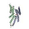 2vkjSC S: Starting model for refinement C: citing same article ( |
|---|---|
| Similar structure data |


-Links
 PDBj
PDBj


- Assembly
| Deposited unit | 
| ||||||||
|---|---|---|---|---|---|---|---|---|---|
| 1 | 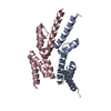
| ||||||||
| 2 | 
| ||||||||
| Unit cell |
|
-Components
| #1: Protein | Mass: 12303.146 Da / Num. of mol.: 4 / Fragment: SOLUBLE DOMAIN, RESIDUES 27-128 Source method: isolated from a genetically manipulated source Source: (gene. exp.) THERMOTOGA MARITIMA (bacteria) / Plasmid: PET28B / Production host: #2: Chemical | ChemComp-PG4 / |   Mass: 194.226 Da / Num. of mol.: 1 / Source method: obtained synthetically / Formula: C8H18O5 / Comment: precipitant*YM Mass: 194.226 Da / Num. of mol.: 1 / Source method: obtained synthetically / Formula: C8H18O5 / Comment: precipitant*YM#3: Chemical | ChemComp-P6G / |   Mass: 282.331 Da / Num. of mol.: 1 / Source method: obtained synthetically / Formula: C12H26O7 / Comment: precipitant*YM Mass: 282.331 Da / Num. of mol.: 1 / Source method: obtained synthetically / Formula: C12H26O7 / Comment: precipitant*YM#4: Water | ChemComp-HOH / |  Mass: 18.015 Da / Num. of mol.: 212 / Source method: isolated from a natural source / Formula: H2O Mass: 18.015 Da / Num. of mol.: 212 / Source method: isolated from a natural source / Formula: H2O |
|---|
-Experimental details
-Experiment
| Experiment | Method: X-RAY DIFFRACTION / Number of used crystals: 1 |
|---|
- Sample preparation
| Crystal | Density Matthews: 2.8 Å3/Da / Density % sol: 54 % / Description: NONE |
|---|---|
| Crystal grow | pH: 5.5 / Details: 50% PEG200, 0.1 M NACITRATE PH 5.5 |
-Data collection
| Diffraction | Mean temperature: 100 K |
|---|---|
| Diffraction source | Source: SYNCHROTRON / Site: ALS  / Beamline: 5.0.1 / Wavelength: 1 / Beamline: 5.0.1 / Wavelength: 1 |
| Detector | Type: ADSC CCD / Detector: CCD / Date: Apr 7, 2006 |
| Radiation | Protocol: SINGLE WAVELENGTH / Monochromatic (M) / Laue (L): M / Scattering type: x-ray |
| Radiation wavelength | Wavelength: 1 Å / Relative weight: 1 |
| Reflection | Resolution: 1.8→50 Å / Num. obs: 51063 / % possible obs: 99.2 % / Observed criterion σ(I): -3 / Redundancy: 3.2 % / Rmerge(I) obs: 0.08 / Net I/σ(I): 17.9 |
| Reflection shell | Resolution: 1.8→1.84 Å / Redundancy: 3.2 % / Rmerge(I) obs: 0.57 / % possible all: 99.6 |
- Processing
| Software |
| ||||||||||||||||||||||||||||||||||||||||||||||||||||||||||||||||||||||||||||||||||||||||||||||||||||||||||||||||||||||||||||||||||||||||||||||||||||||||||||||||||||||||||||||||||||||
|---|---|---|---|---|---|---|---|---|---|---|---|---|---|---|---|---|---|---|---|---|---|---|---|---|---|---|---|---|---|---|---|---|---|---|---|---|---|---|---|---|---|---|---|---|---|---|---|---|---|---|---|---|---|---|---|---|---|---|---|---|---|---|---|---|---|---|---|---|---|---|---|---|---|---|---|---|---|---|---|---|---|---|---|---|---|---|---|---|---|---|---|---|---|---|---|---|---|---|---|---|---|---|---|---|---|---|---|---|---|---|---|---|---|---|---|---|---|---|---|---|---|---|---|---|---|---|---|---|---|---|---|---|---|---|---|---|---|---|---|---|---|---|---|---|---|---|---|---|---|---|---|---|---|---|---|---|---|---|---|---|---|---|---|---|---|---|---|---|---|---|---|---|---|---|---|---|---|---|---|---|---|---|---|
| Refinement | Method to determine structure: MOLECULAR REPLACEMENT Starting model: PDB ENTRY 2VKJ Resolution: 1.79→66.96 Å / Cor.coef. Fo:Fc: 0.926 / Cor.coef. Fo:Fc free: 0.909 / SU B: 6.809 / SU ML: 0.11 / Cross valid method: THROUGHOUT / ESU R: 0.144 / ESU R Free: 0.139 / Stereochemistry target values: MAXIMUM LIKELIHOOD Details: HYDROGENS HAVE BEEN ADDED IN THE RIDING POSITIONS. CHAINS A AND C FORM THE BIOLOGICAL DIMER.
| ||||||||||||||||||||||||||||||||||||||||||||||||||||||||||||||||||||||||||||||||||||||||||||||||||||||||||||||||||||||||||||||||||||||||||||||||||||||||||||||||||||||||||||||||||||||
| Solvent computation | Ion probe radii: 0.8 Å / Shrinkage radii: 0.8 Å / VDW probe radii: 1.4 Å / Solvent model: MASK | ||||||||||||||||||||||||||||||||||||||||||||||||||||||||||||||||||||||||||||||||||||||||||||||||||||||||||||||||||||||||||||||||||||||||||||||||||||||||||||||||||||||||||||||||||||||
| Displacement parameters | Biso mean: 32.58 Å2
| ||||||||||||||||||||||||||||||||||||||||||||||||||||||||||||||||||||||||||||||||||||||||||||||||||||||||||||||||||||||||||||||||||||||||||||||||||||||||||||||||||||||||||||||||||||||
| Refinement step | Cycle: LAST / Resolution: 1.79→66.96 Å
| ||||||||||||||||||||||||||||||||||||||||||||||||||||||||||||||||||||||||||||||||||||||||||||||||||||||||||||||||||||||||||||||||||||||||||||||||||||||||||||||||||||||||||||||||||||||
| Refine LS restraints |
|