 Movie
Movie Controller


+ Open data

- Basic information
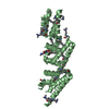
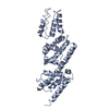
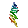
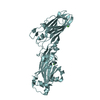
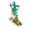
| Entry | Database: PDB / ID: 2vj4 | ||||||
|---|---|---|---|---|---|---|---|
| Title | Methylated Shigella flexneri MxiC | ||||||
 Components Components | PROTEIN MXIC | ||||||
 Keywords Keywords | TRANSPORT PROTEIN / SECRETION REGULATION / T3SS / VIRULENCE / TRANSPORT / TYPE THREE SECRETION SYSTEM | ||||||
| Function / homology |  Function and homology information Function and homology informationprotein secretion by the type III secretion system / outer membrane / negative regulation of protein secretion / host cell / cell surface / extracellular region Similarity search - Function | ||||||
| Biological species |  SHIGELLA FLEXNERI (bacteria) SHIGELLA FLEXNERI (bacteria) | ||||||
| Method |  X-RAY DIFFRACTION / SYNCHROTRON / MOLECULAR REPLACEMENT / Resolution: 2.5 Å X-RAY DIFFRACTION / SYNCHROTRON / MOLECULAR REPLACEMENT / Resolution: 2.5 Å | ||||||
 Authors Authors | Deane, J.E. / Roversi, P. / King, C. / Johnson, S. / Lea, S.M. | ||||||
 Citation Citation | Journal: J.Mol.Biol. / Year: 2008 Title: Structures of the Shigella Flexneri Type 3 Secretion System Protein Mxic Reveal Conformational Variability Amongst Homologues. Authors: Deane, J.E. / Roversi, P. / King, C. / Johnson, S. / Lea, S.M. | ||||||
| History |
|
- Structure visualization
| Structure viewer | Molecule: MolmilJmol/JSmol |
|---|
- Downloads & links
-Download
| PDBx/mmCIF format | 2vj4.cif.gz | 134.6 KB | Display | PDBx/mmCIF format |
|---|---|---|---|---|
| PDB format | pdb2vj4.ent.gz | 107.7 KB | Display | PDB format |
| PDBx/mmJSON format | 2vj4.json.gz | Tree view | PDBx/mmJSON format | |
| Others |  Other downloads Other downloads |
-Validation report
| Arichive directory | https://data.pdbj.org/pub/pdb/validation_reports/vj/2vj4ftp://data.pdbj.org/pub/pdb/validation_reports/vj/2vj4 | HTTPS FTP |
|---|
-Related structure data
| Related structure data |  2vixSC  2vj5C S: Starting model for refinement C: citing same article ( |
|---|---|
| Similar structure data |


-Links
 PDBj
PDBj- Assembly
| Deposited unit | 
| ||||||||
|---|---|---|---|---|---|---|---|---|---|
| 1 | 
| ||||||||
| 2 | 
| ||||||||
| Unit cell |
| ||||||||
| Noncrystallographic symmetry (NCS) | NCS oper: (Code: given Matrix: (-0.00049, 1, 0.00076), Vector: |
-Components
| #1: Protein | Mass: 34184.215 Da / Num. of mol.: 2 / Fragment: RESIDUES 74-355 Source method: isolated from a genetically manipulated source Details: THE CONSTRUCT IS A DELETION OF THE FIRST 73 RESIDUES AND WAS METHYLATED CHEMICALLY FOLLOWING WALTER ET AL, STRUCTURE 14,1617-1622 (2006) Source: (gene. exp.) SHIGELLA FLEXNERI (bacteria) / Strain: PWR100 / Production host: #2: Water | ChemComp-HOH / |  Mass: 18.015 Da / Num. of mol.: 179 / Source method: isolated from a natural source / Formula: H2O Mass: 18.015 Da / Num. of mol.: 179 / Source method: isolated from a natural source / Formula: H2OSequence details | THE CONSTRUCT IS A DELETION OF RESIDUES 1-73 AND THEN HAS THE SEQUENCE HSSGLVPRGSHM FROM THE ...THE CONSTRUCT IS A DELETION OF RESIDUES 1-73 AND THEN HAS THE SEQUENCE HSSGLVPRGS | |
|---|
-Experimental details
-Experiment
| Experiment | Method: X-RAY DIFFRACTION / Number of used crystals: 1 |
|---|
- Sample preparation
| Crystal | Density Matthews: 3.24 Å3/Da / Density % sol: 62 % / Description: NONE |
|---|---|
| Crystal grow | pH: 7 Details: 1.0 M SUCCINIC ACID, 0.1 M HEPES PH 7.0, 1% W/V PEGMME 2000 |
-Data collection
| Diffraction | Mean temperature: 173 K |
|---|---|
| Diffraction source | Source: SYNCHROTRON / Site: ESRF  / Beamline: ID29 / Wavelength: 0.9756 / Beamline: ID29 / Wavelength: 0.9756 |
| Detector | Type: ADSC CCD / Detector: CCD / Date: Jun 22, 2007 |
| Radiation | Protocol: SINGLE WAVELENGTH / Monochromatic (M) / Laue (L): M / Scattering type: x-ray |
| Radiation wavelength | Wavelength: 0.9756 Å / Relative weight: 1 |
| Reflection | Resolution: 2.5→42 Å / Num. obs: 28754 / % possible obs: 99.4 % / Observed criterion σ(I): 0 / Redundancy: 3.5 % / Biso Wilson estimate: 59.3 Å2 / Rmerge(I) obs: 0.07 / Net I/σ(I): 14.2 |
| Reflection shell | Resolution: 2.5→2.64 Å / Redundancy: 3.6 % / Rmerge(I) obs: 0.52 / Mean I/σ(I) obs: 2.7 / % possible all: 99.9 |
- Processing
| Software |
| |||||||||||||||||||||||||||||||||||||||||||||||||||||||||||||||||||||||||||||||||||||||||||||||
|---|---|---|---|---|---|---|---|---|---|---|---|---|---|---|---|---|---|---|---|---|---|---|---|---|---|---|---|---|---|---|---|---|---|---|---|---|---|---|---|---|---|---|---|---|---|---|---|---|---|---|---|---|---|---|---|---|---|---|---|---|---|---|---|---|---|---|---|---|---|---|---|---|---|---|---|---|---|---|---|---|---|---|---|---|---|---|---|---|---|---|---|---|---|---|---|---|
| Refinement | Method to determine structure: MOLECULAR REPLACEMENT Starting model: PDB ENTRY 2VIX Resolution: 2.5→42 Å / Cross valid method: THROUGHOUT / σ(F): 0 Details: REFINED IN BUSTER-TNT VERSION 2.1.1 WITH RESTRAINTS BASED ON REFMAC5 THE LATTICE IS PSEUDOTETRAGONAL P 42 2 2 BUT REFINEMENT IN THIS SPACE GROUP WITH 1 MOL PER ASU DID NOT WORK WELL
| |||||||||||||||||||||||||||||||||||||||||||||||||||||||||||||||||||||||||||||||||||||||||||||||
| Refinement step | Cycle: LAST / Resolution: 2.5→42 Å
| |||||||||||||||||||||||||||||||||||||||||||||||||||||||||||||||||||||||||||||||||||||||||||||||
| Refine LS restraints |
|
