 Movie
Movie Controller


+ Open data

- Basic information
| Entry | Database: PDB / ID: 2vi0 | |||||||||
|---|---|---|---|---|---|---|---|---|---|---|
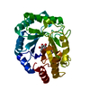
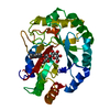
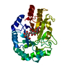
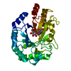
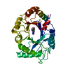
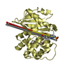
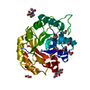
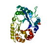
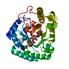
| Title | Lichenase CtLic26 in complex with a thio-oligosaccharide | |||||||||
 Components Components | Endoglucanase H | |||||||||
 Keywords Keywords | HYDROLASE / CARBOHYDRATE METABOLISM / POLYSACCHARIDE DEGRADATION / GLYCOSIDASE / CELLULOSE DEGRADATION / ENZYME GLYCOSIDE HYDROLASE LICHENASE BETA GLUCANASE GH26 G | |||||||||
| Function / homology |  Function and homology information Function and homology informationcellulase / cellulase activity / beta-glucosidase activity / cellulose catabolic process / cell surface / extracellular region Similarity search - Function | |||||||||
| Biological species |  Clostridium thermocellum (bacteria) Clostridium thermocellum (bacteria) | |||||||||
| Method |  X-RAY DIFFRACTION / SYNCHROTRON / MOLECULAR REPLACEMENT / Resolution: 1.51 Å X-RAY DIFFRACTION / SYNCHROTRON / MOLECULAR REPLACEMENT / Resolution: 1.51 Å | |||||||||
 Authors Authors | Money, V.A. / Ducros, V.M. / Davies, G.J. | |||||||||
 Citation Citation | Journal: Org. Biomol. Chem. / Year: 2008 Title: Probing the beta-1,3:1,4 glucanase, CtLic26A, with a thio-oligosaccharide and enzyme variants. Authors: Money, V.A. / Cartmell, A. / Guerreiro, C.I. / Ducros, V.M. / Fontes, C.M. / Gilbert, H.J. / Davies, G.J. #1: Journal: J.Biol.Chem. / Year: 2005Title: How Family 26 Glycoside Hydrolases Orchestrate Catalysis on Different Polysaccharides: Structure and Activity of a Clostridium Thermocellum Lichenase, Ctlic26A. Authors: Taylor, E.J. / Goyal, A. / Guerreiro, C.I.P.D. / Prates, J.A.M. / Money, V.A. / Ferry, N. / Morland, C. / Planas, A. / Macdonald, J.A. / Stick, R.V. / Gilbert, H.J. / Fontes, C.M.G.A. / Davies, G.J. #2: Journal: Angew.Chem.Int.Ed.Engl. / Year: 2006Title: Substrate Distortion by a Lichenase Highlights the Different Conformational Itineraries Harnessed by Related Glycoside Hydrolases. Authors: Money, V.A. / Smith, N.L. / Scaffidi, A. / Stick, R.V. / Gilbert, H.J. / Davies, G.J. | |||||||||
| History |
|
- Structure visualization
| Structure viewer | Molecule: MolmilJmol/JSmol |
|---|
- Downloads & links
-Download
| PDBx/mmCIF format | 2vi0.cif.gz | 145.2 KB | Display | PDBx/mmCIF format |
|---|---|---|---|---|
| PDB format | pdb2vi0.ent.gz | 112.5 KB | Display | PDB format |
| PDBx/mmJSON format | 2vi0.json.gz | Tree view | PDBx/mmJSON format | |
| Others |  Other downloads Other downloads |
-Validation report
| Arichive directory | https://data.pdbj.org/pub/pdb/validation_reports/vi/2vi0ftp://data.pdbj.org/pub/pdb/validation_reports/vi/2vi0 | HTTPS FTP |
|---|
-Related structure data
| Related structure data | 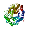 2bv9S  2v80 S: Starting model for refinement |
|---|---|
| Similar structure data |


-Links
 PDBj
PDBj- Assembly
| Deposited unit | 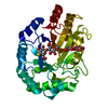
| ||||||||
|---|---|---|---|---|---|---|---|---|---|
| 1 |
| ||||||||
| Unit cell |
|
-Components
| #1: Protein | Mass: 32045.432 Da / Num. of mol.: 1 / Fragment: RESIDUES 26-304 / Mutation: G271E Source method: isolated from a genetically manipulated source Source: (gene. exp.) Clostridium thermocellum (bacteria) / Gene: celH, Cthe_1472 / Production host: | ||
|---|---|---|---|
| #2: Polysaccharide | beta-D-glucopyranose-(1-4)-beta-D-glucopyranose / beta-cellobiose  Source method: isolated from a genetically manipulated source Details: oligosaccharide / References: beta-cellobiose | ||
| #3: Polysaccharide | 4-thio-beta-D-glucopyranose-(1-4)-methyl beta-D-glucopyranoside Source method: isolated from a genetically manipulated source | ||
| #4: Water | ChemComp-HOH /  Mass: 18.015 Da / Num. of mol.: 380 / Source method: isolated from a natural source / Formula: H2O Mass: 18.015 Da / Num. of mol.: 380 / Source method: isolated from a natural source / Formula: H2O | ||
| Compound details | ENGINEERED| Nonpolymer details | METHYL O-BETA-D-GLUCOPYRANOSYL- (1,4)-O-BETA-D-GLUCOPYRAN OSYL)- (1,3)-S-BETA-D-GLUCOPYRANOSYL- ( ...METHYL O-BETA-D-GLUCOPYRAN | |
-Experimental details
-Experiment
| Experiment | Method: X-RAY DIFFRACTION / Number of used crystals: 1 |
|---|
- Sample preparation
| Crystal | Density Matthews: 2.32 Å3/Da / Density % sol: 47 % / Description: NONE |
|---|---|
| Crystal grow | pH: 6.5 Details: 0.15 M AMMONIUM SULPHATE, 30 % PEG 5K, MME, 0.1 M MES PH 6.5 |
-Data collection
| Diffraction | Mean temperature: 100 K |
|---|---|
| Diffraction source | Source: SYNCHROTRON / Site: ESRF  / Beamline: ID14-3 / Wavelength: 0.931 / Beamline: ID14-3 / Wavelength: 0.931 |
| Detector | Type: ADSC CCD / Detector: CCD / Details: MIRRORS |
| Radiation | Monochromator: DIAMOND (111), GE(220) / Protocol: SINGLE WAVELENGTH / Monochromatic (M) / Laue (L): M / Scattering type: x-ray |
| Radiation wavelength | Wavelength: 0.931 Å / Relative weight: 1 |
| Reflection | Resolution: 1.51→34.88 Å / Num. obs: 3620 / % possible obs: 95.8 % / Redundancy: 5.1 % / Rmerge(I) obs: 0.09 / Net I/σ(I): 11.8 |
| Reflection shell | Resolution: 1.51→1.6 Å / Redundancy: 3 % / Rmerge(I) obs: 0.5 / Mean I/σ(I) obs: 2 / % possible all: 72 |
- Processing
| Software |
| ||||||||||||||||||||||||||||||||||||||||||||||||||||||||||||||||||||||||||||||||||||||||||||||||||||||||||||||||||||||||||||||||||||||||||||||||||||||||||||||||||||||||||||||||||||||
|---|---|---|---|---|---|---|---|---|---|---|---|---|---|---|---|---|---|---|---|---|---|---|---|---|---|---|---|---|---|---|---|---|---|---|---|---|---|---|---|---|---|---|---|---|---|---|---|---|---|---|---|---|---|---|---|---|---|---|---|---|---|---|---|---|---|---|---|---|---|---|---|---|---|---|---|---|---|---|---|---|---|---|---|---|---|---|---|---|---|---|---|---|---|---|---|---|---|---|---|---|---|---|---|---|---|---|---|---|---|---|---|---|---|---|---|---|---|---|---|---|---|---|---|---|---|---|---|---|---|---|---|---|---|---|---|---|---|---|---|---|---|---|---|---|---|---|---|---|---|---|---|---|---|---|---|---|---|---|---|---|---|---|---|---|---|---|---|---|---|---|---|---|---|---|---|---|---|---|---|---|---|---|---|
| Refinement | Method to determine structure: MOLECULAR REPLACEMENT Starting model: PDB ENTRY 2BV9 Resolution: 1.51→34.88 Å / Cor.coef. Fo:Fc: 0.966 / Cor.coef. Fo:Fc free: 0.953 / SU B: 3.274 / SU ML: 0.055 / Cross valid method: THROUGHOUT / ESU R: 0.117 / ESU R Free: 0.081 / Stereochemistry target values: MAXIMUM LIKELIHOOD / Details: HYDROGENS HAVE BEEN ADDED IN THE RIDING POSITIONS.
| ||||||||||||||||||||||||||||||||||||||||||||||||||||||||||||||||||||||||||||||||||||||||||||||||||||||||||||||||||||||||||||||||||||||||||||||||||||||||||||||||||||||||||||||||||||||
| Solvent computation | Ion probe radii: 0.8 Å / Shrinkage radii: 0.8 Å / VDW probe radii: 1.2 Å / Solvent model: MASK | ||||||||||||||||||||||||||||||||||||||||||||||||||||||||||||||||||||||||||||||||||||||||||||||||||||||||||||||||||||||||||||||||||||||||||||||||||||||||||||||||||||||||||||||||||||||
| Displacement parameters | Biso mean: 9.22 Å2
| ||||||||||||||||||||||||||||||||||||||||||||||||||||||||||||||||||||||||||||||||||||||||||||||||||||||||||||||||||||||||||||||||||||||||||||||||||||||||||||||||||||||||||||||||||||||
| Refinement step | Cycle: LAST / Resolution: 1.51→34.88 Å
| ||||||||||||||||||||||||||||||||||||||||||||||||||||||||||||||||||||||||||||||||||||||||||||||||||||||||||||||||||||||||||||||||||||||||||||||||||||||||||||||||||||||||||||||||||||||
| Refine LS restraints |
|