Protocol: SINGLE WAVELENGTH / Monochromatic (M) / Laue (L): M / Scattering type: x-ray
Radiation wavelength
Wavelength: 0.873 Å / Relative weight: 1
Reflection
Resolution: 2.8→60 Å / Num. obs: 43922 / % possible obs: 95.6 % / Observed criterion σ(I): 2 / Redundancy: 2.7 % / Rmerge(I) obs: 0.12 / Net I/σ(I): 9.2
Reflection shell
Resolution: 2.8→2.95 Å / Redundancy: 2.8 % / Rmerge(I) obs: 0.46 / Mean I/σ(I) obs: 2.3 / % possible all: 94.4
-
Processing
Software
Name
Version
Classification
REFMAC
5.2.0019
refinement
MOSFLM
datareduction
SCALA
datascaling
MOLREP
phasing
Refinement
Method to determine structure: MOLECULAR REPLACEMENT / Resolution: 2.8→35 Å / Cor.coef. Fo:Fc: 0.908 / Cor.coef. Fo:Fc free: 0.877 / SU B: 14.178 / SU ML: 0.279 / Cross valid method: THROUGHOUT / ESU R Free: 0.395 / Stereochemistry target values: MAXIMUM LIKELIHOOD / Details: HYDROGENS HAVE BEEN ADDED IN THE RIDING POSITIONS
Rfactor
Num. reflection
% reflection
Selection details
Rfree
0.255
2237
5.1 %
RANDOM
Rwork
0.215
-
-
-
obs
0.217
41665
94.8 %
-
Solvent computation
Ion probe radii: 0.8 Å / Shrinkage radii: 0.8 Å / VDW probe radii: 1.2 Å / Solvent model: MASK
 Movie
Movie Controller

 Yorodumi
 Open data
 Basic information
 Components
 Keywords
 Function and homology information
 X-RAY DIFFRACTION /
  Authors
 Citation
 Structure visualization
 Downloads & links
 Other downloads
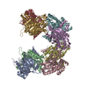


 PDBj
PDBj Assembly
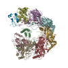
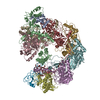
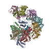


 Sample preparation
 / Beamline: ID23-2 / Wavelength: 0.873
  Processing