 Movie
Movie Controller

[English] 日本語
 Yorodumi
- PDB-2v7s: Crystal structure of the putative lipoprotein LppA from Mycobacte... -
+ Open data

- Basic information
| Entry | Database: PDB / ID: 2v7s | ||||||
|---|---|---|---|---|---|---|---|
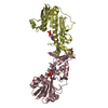
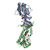
| Title | Crystal structure of the putative lipoprotein LppA from Mycobacterium tuberculosis | ||||||
 Components Components | PROBABLE CONSERVED LIPOPROTEIN LPPA | ||||||
 Keywords Keywords | UNKNOWN FUNCTION / LIPOPROTEIN / PUTATIVE LIPOPROTEIN | ||||||
| Function / homology | TBP-like - #20 / Putative lipoprotein, pathogenic bacteria / Lipoprotein confined to pathogenic Mycobacterium / TBP-like / 2-Layer Sandwich / plasma membrane / Alpha Beta / Putative lipoprotein LppA / Putative lipoprotein LppA Function and homology information Function and homology information | ||||||
| Biological species |   MYCOBACTERIUM TUBERCULOSIS (bacteria) MYCOBACTERIUM TUBERCULOSIS (bacteria) | ||||||
| Method |  X-RAY DIFFRACTION / SYNCHROTRON / SAD / Resolution: 1.96 Å X-RAY DIFFRACTION / SYNCHROTRON / SAD / Resolution: 1.96 Å | ||||||
 Authors Authors | Grana, M. / Miras, I. / Haouz, A. / Winter, N. / Buschiazzo, A. / Bellinzoni, M. / Alzari, P.M. | ||||||
 Citation Citation | Journal: Proteins / Year: 2010 Title: Crystal Structure of Mycobacterium Tuberculosis Lppa, a Lipoprotein Confined to Pathogenic Mycobacteria. Authors: Grana, M. / Bellinzoni, M. / Bellalou, J. / Haouz, A. / Miras, I. / Buschiazzo, A. / Winter, N. / Alzari, P.M. | ||||||
| History |
|
- Structure visualization
| Structure viewer | Molecule: MolmilJmol/JSmol |
|---|
- Downloads & links
-Download
| PDBx/mmCIF format | 2v7s.cif.gz | 49 KB | Display | PDBx/mmCIF format |
|---|---|---|---|---|
| PDB format | pdb2v7s.ent.gz | 34 KB | Display | PDB format |
| PDBx/mmJSON format | 2v7s.json.gz | Tree view | PDBx/mmJSON format | |
| Others |  Other downloads Other downloads |
-Validation report
| Arichive directory | https://data.pdbj.org/pub/pdb/validation_reports/v7/2v7sftp://data.pdbj.org/pub/pdb/validation_reports/v7/2v7s | HTTPS FTP |
|---|
-Related structure data
| Similar structure data |
|---|


-Links
 PDBj
PDBj- Assembly
| Deposited unit | 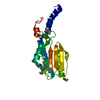
| ||||||||
|---|---|---|---|---|---|---|---|---|---|
| 1 | 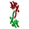
| ||||||||
| Unit cell |
|
-Components
| #1: Protein | Mass: 24337.549 Da / Num. of mol.: 1 / Fragment: RESIDUES 34-219 Source method: isolated from a genetically manipulated source Source: (gene. exp.) MYCOBACTERIUM TUBERCULOSIS (bacteria) / Strain: H37RV / Production host: | ||||||
|---|---|---|---|---|---|---|---|
| #2: Chemical |   Mass: 92.094 Da / Num. of mol.: 3 / Source method: obtained synthetically / Formula: C3H8O3 Mass: 92.094 Da / Num. of mol.: 3 / Source method: obtained synthetically / Formula: C3H8O3#3: Water | ChemComp-HOH / |  Mass: 18.015 Da / Num. of mol.: 62 / Source method: isolated from a natural source / Formula: H2O Mass: 18.015 Da / Num. of mol.: 62 / Source method: isolated from a natural source / Formula: H2OHas protein modification | Y | Sequence details | THE RECOMBINANT PROTEIN INCLUDES A N-TERMINAL TAG ( SEQUENCE MSYYHHHHHHLESTSLYKKAGSENLYFQG) FUSED ...THE RECOMBINAN | |
-Experimental details
-Experiment
| Experiment | Method: X-RAY DIFFRACTION / Number of used crystals: 1 |
|---|
- Sample preparation
| Crystal | Density Matthews: 2.81 Å3/Da / Density % sol: 56 % / Description: NONE |
|---|---|
| Crystal grow | pH: 9 / Details: 20% PEG-3350, BICINE 0.1 M, PH 9, 50 MM MGCL2 |
-Data collection
| Diffraction | Mean temperature: 100 K |
|---|---|
| Diffraction source | Source: SYNCHROTRON / Site: ESRF  / Beamline: ID14-4 / Wavelength: 0.979 / Beamline: ID14-4 / Wavelength: 0.979 |
| Detector | Type: ADSC CCD / Detector: CCD / Date: Mar 21, 2006 |
| Radiation | Protocol: SINGLE WAVELENGTH / Monochromatic (M) / Laue (L): M / Scattering type: x-ray |
| Radiation wavelength | Wavelength: 0.979 Å / Relative weight: 1 |
| Reflection | Resolution: 1.96→45 Å / Num. obs: 15229 / % possible obs: 97.8 % / Observed criterion σ(I): 0 / Redundancy: 3.9 % / Rmerge(I) obs: 0.11 / Net I/σ(I): 8.6 |
| Reflection shell | Resolution: 1.96→2.07 Å / Redundancy: 3.1 % / Rmerge(I) obs: 0.32 / Mean I/σ(I) obs: 3.7 / % possible all: 85.6 |
- Processing
| Software |
| ||||||||||||||||||||||||||||||||||||||||||||||||||||||||||||||||||||||||||||||||||||||||||||||||||||||||||||||||||||||||||||||||||||||||||||||||||||||||||||||||||||||||||||||||||||||
|---|---|---|---|---|---|---|---|---|---|---|---|---|---|---|---|---|---|---|---|---|---|---|---|---|---|---|---|---|---|---|---|---|---|---|---|---|---|---|---|---|---|---|---|---|---|---|---|---|---|---|---|---|---|---|---|---|---|---|---|---|---|---|---|---|---|---|---|---|---|---|---|---|---|---|---|---|---|---|---|---|---|---|---|---|---|---|---|---|---|---|---|---|---|---|---|---|---|---|---|---|---|---|---|---|---|---|---|---|---|---|---|---|---|---|---|---|---|---|---|---|---|---|---|---|---|---|---|---|---|---|---|---|---|---|---|---|---|---|---|---|---|---|---|---|---|---|---|---|---|---|---|---|---|---|---|---|---|---|---|---|---|---|---|---|---|---|---|---|---|---|---|---|---|---|---|---|---|---|---|---|---|---|---|
| Refinement | Method to determine structure: SAD Starting model: NONE Resolution: 1.96→6 Å / Cor.coef. Fo:Fc: 0.939 / Cor.coef. Fo:Fc free: 0.912 / SU B: 5.91 / SU ML: 0.086 / TLS residual ADP flag: LIKELY RESIDUAL / Cross valid method: THROUGHOUT / ESU R: 0.155 / ESU R Free: 0.141 / Stereochemistry target values: MAXIMUM LIKELIHOOD / Details: HYDROGENS HAVE BEEN ADDED IN THE RIDING POSITIONS.
| ||||||||||||||||||||||||||||||||||||||||||||||||||||||||||||||||||||||||||||||||||||||||||||||||||||||||||||||||||||||||||||||||||||||||||||||||||||||||||||||||||||||||||||||||||||||
| Solvent computation | Ion probe radii: 0.8 Å / Shrinkage radii: 0.8 Å / VDW probe radii: 1.2 Å / Solvent model: MASK | ||||||||||||||||||||||||||||||||||||||||||||||||||||||||||||||||||||||||||||||||||||||||||||||||||||||||||||||||||||||||||||||||||||||||||||||||||||||||||||||||||||||||||||||||||||||
| Displacement parameters | Biso mean: 23.93 Å2
| ||||||||||||||||||||||||||||||||||||||||||||||||||||||||||||||||||||||||||||||||||||||||||||||||||||||||||||||||||||||||||||||||||||||||||||||||||||||||||||||||||||||||||||||||||||||
| Refinement step | Cycle: LAST / Resolution: 1.96→6 Å
| ||||||||||||||||||||||||||||||||||||||||||||||||||||||||||||||||||||||||||||||||||||||||||||||||||||||||||||||||||||||||||||||||||||||||||||||||||||||||||||||||||||||||||||||||||||||
| Refine LS restraints |
|