 Movie
Movie Controller


+ Open data

- Basic information
| Entry | Database: PDB / ID: 2v5w | ||||||
|---|---|---|---|---|---|---|---|
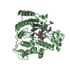
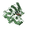
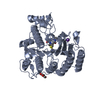
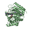
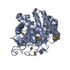
| Title | Crystal structure of HDAC8-substrate complex | ||||||
 Components Components |
| ||||||
 Keywords Keywords | HYDROLASE/HYDROLASE SUBSTRATE / HISTONE DEACETYLASE / CHROMATIN REGULATOR / P53 / HDAC / HDAC8 / NUCLEUS / REPRESSOR / HYDROLASE / NUCLEAR PROTEIN / PEPTIDIC SUBSTRATE / TRANSCRIPTION REGULATION / CHROMATIN / TRANSCRIPTION / DEACETYLATION / HYDROLASE-HYDROLASE SUBSTRATE COMPLEX | ||||||
| Function / homology |  Function and homology information Function and homology informationhistone decrotonylase activity / histone deacetylase activity, hydrolytic mechanism / histone deacetylase / negative regulation of helicase activity / Loss of function of TP53 in cancer due to loss of tetramerization ability / Regulation of TP53 Expression / signal transduction by p53 class mediator / negative regulation of G1 to G0 transition / negative regulation of glucose catabolic process to lactate via pyruvate / Transcriptional activation of cell cycle inhibitor p21 ...histone decrotonylase activity / histone deacetylase activity, hydrolytic mechanism / histone deacetylase / negative regulation of helicase activity / Loss of function of TP53 in cancer due to loss of tetramerization ability / Regulation of TP53 Expression / signal transduction by p53 class mediator / negative regulation of G1 to G0 transition / negative regulation of glucose catabolic process to lactate via pyruvate / Transcriptional activation of cell cycle inhibitor p21 / regulation of intrinsic apoptotic signaling pathway by p53 class mediator / negative regulation of pentose-phosphate shunt / ATP-dependent DNA/DNA annealing activity / Activation of NOXA and translocation to mitochondria / regulation of cell cycle G2/M phase transition / regulation of fibroblast apoptotic process / intrinsic apoptotic signaling pathway in response to hypoxia / oligodendrocyte apoptotic process / negative regulation of miRNA processing / positive regulation of thymocyte apoptotic process / oxidative stress-induced premature senescence / regulation of tissue remodeling / positive regulation of mitochondrial membrane permeability / mRNA transcription / positive regulation of programmed necrotic cell death / bone marrow development / circadian behavior / T cell proliferation involved in immune response / regulation of mitochondrial membrane permeability involved in apoptotic process / germ cell nucleus / regulation of telomere maintenance / RUNX3 regulates CDKN1A transcription / protein lysine deacetylase activity / glucose catabolic process to lactate via pyruvate / Hydrolases; Acting on carbon-nitrogen bonds, other than peptide bonds; In linear amides / TP53 Regulates Transcription of Death Receptors and Ligands / TP53 regulates transcription of additional cell cycle genes whose exact role in the p53 pathway remain uncertain / Activation of PUMA and translocation to mitochondria / regulation of DNA damage response, signal transduction by p53 class mediator / histone deacetylase regulator activity / negative regulation of glial cell proliferation / Regulation of TP53 Activity through Association with Co-factors / mitotic sister chromatid cohesion / negative regulation of neuroblast proliferation / mitochondrial DNA repair / T cell lineage commitment / histone deacetylase activity / Formation of Senescence-Associated Heterochromatin Foci (SAHF) / nuclear chromosome / ER overload response / thymocyte apoptotic process / B cell lineage commitment / TP53 Regulates Transcription of Caspase Activators and Caspases / Notch-HLH transcription pathway / negative regulation of mitophagy / cardiac septum morphogenesis / negative regulation of DNA replication / entrainment of circadian clock by photoperiod / negative regulation of telomere maintenance via telomerase / Zygotic genome activation (ZGA) / PI5P Regulates TP53 Acetylation / positive regulation of release of cytochrome c from mitochondria / Association of TriC/CCT with target proteins during biosynthesis / necroptotic process / TP53 Regulates Transcription of Genes Involved in Cytochrome C Release / SUMOylation of transcription factors / TP53 regulates transcription of several additional cell death genes whose specific roles in p53-dependent apoptosis remain uncertain / TFIID-class transcription factor complex binding / intrinsic apoptotic signaling pathway by p53 class mediator / negative regulation of reactive oxygen species metabolic process / rRNA transcription / histone deacetylase complex / Transcriptional Regulation by VENTX / replicative senescence / general transcription initiation factor binding / cellular response to UV-C / cellular response to actinomycin D / intrinsic apoptotic signaling pathway in response to endoplasmic reticulum stress / positive regulation of execution phase of apoptosis / positive regulation of RNA polymerase II transcription preinitiation complex assembly / neuroblast proliferation / intrinsic apoptotic signaling pathway in response to DNA damage by p53 class mediator / response to X-ray / hematopoietic stem cell differentiation / Pyroptosis / viral process / embryonic organ development / chromosome organization / type II interferon-mediated signaling pathway / TP53 Regulates Transcription of Genes Involved in G1 Cell Cycle Arrest / somitogenesis / hematopoietic progenitor cell differentiation / glial cell proliferation / negative regulation of fibroblast proliferation / core promoter sequence-specific DNA binding / positive regulation of cardiac muscle cell apoptotic process / negative regulation of stem cell proliferation / cellular response to glucose starvation / mitophagy / cis-regulatory region sequence-specific DNA binding Similarity search - Function | ||||||
| Biological species |  HOMO SAPIENS (human) HOMO SAPIENS (human)SYNTHETIC CONSTRUCT (others) | ||||||
| Method |  X-RAY DIFFRACTION / SYNCHROTRON / MOLECULAR REPLACEMENT / Resolution: 2 Å X-RAY DIFFRACTION / SYNCHROTRON / MOLECULAR REPLACEMENT / Resolution: 2 Å | ||||||
 Authors Authors | Di Marco, S. / Vannini, A. / Volpari, C. | ||||||
 Citation Citation | Journal: Embo Rep. / Year: 2007 Title: Substrate Binding to Histone Deacetylases as Revealed by Crystal Structure of Hdac8-Substrate Complex Authors: Vannini, A. / Volpari, C. / Gallinari, P. / Jones, P. / Mattu, M. / Carfi, A. / Defrancesco, R. / Steinkuhler, C. / Di Marco, S. | ||||||
| History |
|
- Structure visualization
| Structure viewer | Molecule: MolmilJmol/JSmol |
|---|
- Downloads & links
-Download
| PDBx/mmCIF format | 2v5w.cif.gz | 174.2 KB | Display | PDBx/mmCIF format |
|---|---|---|---|---|
| PDB format | pdb2v5w.ent.gz | 136.7 KB | Display | PDB format |
| PDBx/mmJSON format | 2v5w.json.gz | Tree view | PDBx/mmJSON format | |
| Others |  Other downloads Other downloads |
-Validation report
| Summary document | 2v5w_validation.pdf.gz | 463.9 KB | Display | wwPDB validaton report |
|---|---|---|---|---|
| Full document | 2v5w_full_validation.pdf.gz | 472.5 KB | Display | |
| Data in XML | 2v5w_validation.xml.gz | 36.1 KB | Display | |
| Data in CIF | 2v5w_validation.cif.gz | 55.3 KB | Display | |
| Arichive directory | https://data.pdbj.org/pub/pdb/validation_reports/v5/2v5wftp://data.pdbj.org/pub/pdb/validation_reports/v5/2v5w | HTTPS FTP |
-Related structure data
| Related structure data |  2v5xC  1w22S C: citing same article ( S: Starting model for refinement |
|---|---|
| Similar structure data |


-Links
 PDBj
PDBj


- Assembly
| Deposited unit | 
| ||||||||
|---|---|---|---|---|---|---|---|---|---|
| 1 | 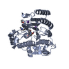
| ||||||||
| 2 | 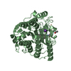
| ||||||||
| Unit cell |
| ||||||||
| Noncrystallographic symmetry (NCS) | NCS oper: (Code: given Matrix: (-1, 0.00146, 0.00018), Vector: |
-Components
-Protein , 1 types, 2 molecules AB
| #1: Protein | Mass: 43158.941 Da / Num. of mol.: 2 / Mutation: YES Source method: isolated from a genetically manipulated source Source: (gene. exp.) HOMO SAPIENS (human) / Plasmid: PET21B / Production host:  |
|---|
-Protein/peptide , 2 types, 3 molecules GIL
| #2: Protein/peptide | Mass: 189.171 Da / Num. of mol.: 1 / Source method: obtained synthetically / Source: (synth.) SYNTHETIC CONSTRUCT (others) |
|---|---|
| #3: Protein/peptide | Mass: 836.980 Da / Num. of mol.: 2 / Source method: obtained synthetically / Source: (synth.) SYNTHETIC CONSTRUCT (others) / References: UniProt: P04637*PLUS |
-Non-polymers , 3 types, 752 molecules 


| #4: Chemical | ChemComp-K /  Mass: 39.098 Da / Num. of mol.: 4 / Source method: obtained synthetically / Formula: K Mass: 39.098 Da / Num. of mol.: 4 / Source method: obtained synthetically / Formula: K#5: Chemical |  Mass: 65.409 Da / Num. of mol.: 2 / Source method: obtained synthetically / Formula: Zn Mass: 65.409 Da / Num. of mol.: 2 / Source method: obtained synthetically / Formula: Zn#6: Water | ChemComp-HOH / | Mass: 18.015 Da / Num. of mol.: 746 / Source method: isolated from a natural source / Formula: H2O |
|---|
-Details
| Compound details | ENGINEERED| Has protein modification | Y | Sequence details | Y306 MUTATED TO F306. IEGRSHHHHH | |
|---|
-Experimental details
-Experiment
| Experiment | Method: X-RAY DIFFRACTION |
|---|
- Sample preparation
| Crystal | Density Matthews: 2.21 Å3/Da / Density % sol: 43.95 % / Description: NONE |
|---|---|
| Crystal grow | Method: vapor diffusion, hanging drop Details: HDAC8 POINT MUTANT Y306F, IN 50 MM TRIS-HCL PH 8.0, 5% GLYCEROL, 1 MM DTT, 150 MM KCL, WAS CONCENTRATED TO 217 UM, RESPECTIVELY. Y306F-HDAC8 PLUS 15 MOLAR EXCESSES OF SUBSTRATE, WAS ...Details: HDAC8 POINT MUTANT Y306F, IN 50 MM TRIS-HCL PH 8.0, 5% GLYCEROL, 1 MM DTT, 150 MM KCL, WAS CONCENTRATED TO 217 UM, RESPECTIVELY. Y306F-HDAC8 PLUS 15 MOLAR EXCESSES OF SUBSTRATE, WAS CRYSTALLIZED AT RT BY THE HANGING-DROP METHOD IN 50 MM TRIS-HCL PH 8.0, 50 MM MGCL2, 10% PEG 4,000, 2 MM TRI(2-CARBOXYETHYL)PHOSPHIN (TCEP) AND 30 MM GLYCYL-GLYCYL- GLYCINE. |
-Data collection
| Diffraction | Mean temperature: 100 K |
|---|---|
| Diffraction source | Source: SYNCHROTRON / Site: ESRF  / Beamline: ID14-3 / Wavelength: 0.931 / Beamline: ID14-3 / Wavelength: 0.931 |
| Detector | Type: ADSC CCD / Detector: CCD |
| Radiation | Protocol: SINGLE WAVELENGTH / Monochromatic (M) / Laue (L): M / Scattering type: x-ray |
| Radiation wavelength | Wavelength: 0.931 Å / Relative weight: 1 |
| Reflection | Resolution: 2→50 Å / Num. obs: 53718 / % possible obs: 95.6 % / Observed criterion σ(I): 3 / Redundancy: 4.8 % / Rmerge(I) obs: 0.05 / Net I/σ(I): 18.2 |
| Reflection shell | Resolution: 2→2.06 Å / Redundancy: 2 % / Rmerge(I) obs: 0.15 / Mean I/σ(I) obs: 6.1 / % possible all: 59.4 |
- Processing
| Software |
| ||||||||||||||||||||||||||||||||||||||||||||||||||||||||||||||||||||||||||||||||||||||||||||||||||||||||||||||||||||||||||||||||||||||||||||||||||||||||||||||||||||||||||||||||||||||
|---|---|---|---|---|---|---|---|---|---|---|---|---|---|---|---|---|---|---|---|---|---|---|---|---|---|---|---|---|---|---|---|---|---|---|---|---|---|---|---|---|---|---|---|---|---|---|---|---|---|---|---|---|---|---|---|---|---|---|---|---|---|---|---|---|---|---|---|---|---|---|---|---|---|---|---|---|---|---|---|---|---|---|---|---|---|---|---|---|---|---|---|---|---|---|---|---|---|---|---|---|---|---|---|---|---|---|---|---|---|---|---|---|---|---|---|---|---|---|---|---|---|---|---|---|---|---|---|---|---|---|---|---|---|---|---|---|---|---|---|---|---|---|---|---|---|---|---|---|---|---|---|---|---|---|---|---|---|---|---|---|---|---|---|---|---|---|---|---|---|---|---|---|---|---|---|---|---|---|---|---|---|---|---|
| Refinement | Method to determine structure: MOLECULAR REPLACEMENT Starting model: PDB ENTRY 1W22 Resolution: 2→50 Å / Cor.coef. Fo:Fc: 0.943 / Cor.coef. Fo:Fc free: 0.915 / SU B: 4.232 / SU ML: 0.12 / Cross valid method: THROUGHOUT / ESU R: 0.192 / ESU R Free: 0.172 / Stereochemistry target values: MAXIMUM LIKELIHOOD / Details: HYDROGENS HAVE BEEN ADDED IN THE RIDING POSITIONS.
| ||||||||||||||||||||||||||||||||||||||||||||||||||||||||||||||||||||||||||||||||||||||||||||||||||||||||||||||||||||||||||||||||||||||||||||||||||||||||||||||||||||||||||||||||||||||
| Solvent computation | Ion probe radii: 0.8 Å / Shrinkage radii: 0.8 Å / VDW probe radii: 1.4 Å / Solvent model: BABINET MODEL WITH MASK | ||||||||||||||||||||||||||||||||||||||||||||||||||||||||||||||||||||||||||||||||||||||||||||||||||||||||||||||||||||||||||||||||||||||||||||||||||||||||||||||||||||||||||||||||||||||
| Displacement parameters | Biso mean: 18.7 Å2
| ||||||||||||||||||||||||||||||||||||||||||||||||||||||||||||||||||||||||||||||||||||||||||||||||||||||||||||||||||||||||||||||||||||||||||||||||||||||||||||||||||||||||||||||||||||||
| Refinement step | Cycle: LAST / Resolution: 2→50 Å
| ||||||||||||||||||||||||||||||||||||||||||||||||||||||||||||||||||||||||||||||||||||||||||||||||||||||||||||||||||||||||||||||||||||||||||||||||||||||||||||||||||||||||||||||||||||||
| Refine LS restraints |
|