 Movie
Movie Controller


+ Open data

- Basic information
| Entry | Database: PDB / ID: 2v5r | ||||||
|---|---|---|---|---|---|---|---|
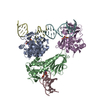
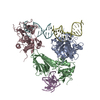
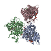
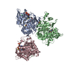
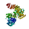
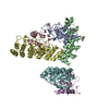
| Title | Structural basis for Dscam isoform specificity | ||||||
 Components Components | DSCAM | ||||||
 Keywords Keywords | CELL ADHESION / DOWN SYNDROME CELL ADHESION MOLECULE DSCAM NEUROBIOLOGY SPL IMMUNOGLOBULIN DOMAIN / MEMBRANE / DEVELOPMENTAL PROTEIN | ||||||
| Function / homology |  Function and homology information Function and homology informationDSCAM interactions / mushroom body development / detection of molecule of bacterial origin / central nervous system morphogenesis / ventral cord development / detection of mechanical stimulus involved in sensory perception of touch / axon guidance receptor activity / axon extension involved in axon guidance / dendrite self-avoidance / peripheral nervous system development ...DSCAM interactions / mushroom body development / detection of molecule of bacterial origin / central nervous system morphogenesis / ventral cord development / detection of mechanical stimulus involved in sensory perception of touch / axon guidance receptor activity / axon extension involved in axon guidance / dendrite self-avoidance / peripheral nervous system development / axonal fasciculation / regulation of axonogenesis / regulation of dendrite morphogenesis / antigen binding / neuron development / phagocytosis / axon guidance / perikaryon / cell adhesion / neuron projection / axon / neuronal cell body / dendrite / extracellular region / identical protein binding / plasma membrane Similarity search - Function | ||||||
| Biological species |  | ||||||
| Method |  X-RAY DIFFRACTION / SYNCHROTRON / MOLECULAR REPLACEMENT / Resolution: 3 Å X-RAY DIFFRACTION / SYNCHROTRON / MOLECULAR REPLACEMENT / Resolution: 3 Å | ||||||
 Authors Authors | Meijers, R. / Puettmann-Holgado, R. / Skiniotis, G. / Liu, J.-H. / Walz, T. / Schmucker, D. / Wang, J.-H. | ||||||
 Citation Citation | Journal: Nature / Year: 2007 Title: Structural Basis of Dscam Isoform Specificity Authors: Meijers, R. / Puettmann-Holgado, R. / Skiniotis, G. / Liu, J.-H. / Walz, T. / Wang, J.-H. / Schmucker, D. | ||||||
| History |
| ||||||
| Remark 700 | SHEET THE SHEET STRUCTURE OF THIS MOLECULE IS BIFURCATED. IN ORDER TO REPRESENT THIS FEATURE IN ... SHEET THE SHEET STRUCTURE OF THIS MOLECULE IS BIFURCATED. IN ORDER TO REPRESENT THIS FEATURE IN THE SHEET RECORDS BELOW, TWO SHEETS ARE DEFINED. |
- Structure visualization
| Structure viewer | Molecule: MolmilJmol/JSmol |
|---|
- Downloads & links
-Download
| PDBx/mmCIF format | 2v5r.cif.gz | 150.1 KB | Display | PDBx/mmCIF format |
|---|---|---|---|---|
| PDB format | pdb2v5r.ent.gz | 121.1 KB | Display | PDB format |
| PDBx/mmJSON format | 2v5r.json.gz | Tree view | PDBx/mmJSON format | |
| Others |  Other downloads Other downloads |
-Validation report
| Arichive directory | https://data.pdbj.org/pub/pdb/validation_reports/v5/2v5rftp://data.pdbj.org/pub/pdb/validation_reports/v5/2v5r | HTTPS FTP |
|---|
-Related structure data
| Related structure data | 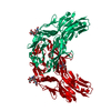 2v5mSC  2v5sC S: Starting model for refinement C: citing same article ( |
|---|---|
| Similar structure data |


-Links
 PDBj
PDBj


- Assembly
| Deposited unit | 
| ||||||||
|---|---|---|---|---|---|---|---|---|---|
| 1 |
| ||||||||
| Unit cell |
| ||||||||
| Noncrystallographic symmetry (NCS) | NCS oper: (Code: given Matrix: (0.863, -0.001, 0.506), Vector: Details | THE DIMERIC STATE DESCRIBED IN REMARK 350 BELOW HASHAS BEEN EXPERIMENTALLY VALIDATED USIING BEAD AGGREGATIONASSAYS. | |
-Components
| #1: Protein | Mass: 43476.008 Da / Num. of mol.: 2 Fragment: N-TERMINAL FOUR DOMAINS (D1, D2, D3 AND D4), RESIDUES 36-423 Source method: isolated from a genetically manipulated source Details: ISOFORM 4.9/6.9 / Source: (gene. exp.)  SPODOPTERA FRUGIPERDA (fall armyworm) / References: UniProt: Q9NBA1, UniProt: Q0E9K4*PLUS SPODOPTERA FRUGIPERDA (fall armyworm) / References: UniProt: Q9NBA1, UniProt: Q0E9K4*PLUS#2: Sugar | ChemComp-NAG /   Type: D-saccharide, beta linking / Mass: 221.208 Da / Num. of mol.: 8 Type: D-saccharide, beta linking / Mass: 221.208 Da / Num. of mol.: 8Source method: isolated from a genetically manipulated source Formula: C8H15NO6 #3: Chemical |   Mass: 92.094 Da / Num. of mol.: 2 / Source method: obtained synthetically / Formula: C3H8O3 Mass: 92.094 Da / Num. of mol.: 2 / Source method: obtained synthetically / Formula: C3H8O3Has protein modification | Y | Sequence details | THE CONFLICTS GIVEN IN THE SEQADV RECORDS BELOW ARE AS A RESULT OF A SPLICE VARIANT FORM OF THE ...THE CONFLICTS GIVEN IN THE SEQADV RECORDS BELOW ARE AS A RESULT OF A SPLICE VARIANT FORM OF THE PROTEIN WHERE EXON 4 COVERING RESIDUES 102 TO 156 CONSISTS OF ISOFORM 9 AND EXON 6 COVERING RESIDUES 205 TO 245 CONSISTS OF ISOFORM 9. | |
|---|
-Experimental details
-Experiment
| Experiment | Method: X-RAY DIFFRACTION / Number of used crystals: 1 |
|---|
- Sample preparation
| Crystal | Density Matthews: 3.9 Å3/Da / Density % sol: 69 % / Description: NONE |
|---|---|
| Crystal grow | Details: 10 % PEG 8000, 1MM SPERMIDINE, 0.1 M TRISHCL PH 8.5 |
-Data collection
| Diffraction | Mean temperature: 100 K |
|---|---|
| Diffraction source | Source: SYNCHROTRON / Site: APS  / Beamline: 19-ID / Wavelength: 0.979 / Beamline: 19-ID / Wavelength: 0.979 |
| Detector | Type: ADSC CCD / Detector: CCD / Date: Jul 9, 2005 |
| Radiation | Protocol: SINGLE WAVELENGTH / Monochromatic (M) / Laue (L): M / Scattering type: x-ray |
| Radiation wavelength | Wavelength: 0.979 Å / Relative weight: 1 |
| Reflection | Resolution: 3→20 Å / Num. obs: 53580 / % possible obs: 86 % / Observed criterion σ(I): 0 / Redundancy: 2.8 % / Rmerge(I) obs: 0.1 / Net I/σ(I): 10.9 |
| Reflection shell | Resolution: 3→3.2 Å / Rmerge(I) obs: 0.23 / Mean I/σ(I) obs: 1.9 / % possible all: 55.5 |
- Processing
| Software |
| ||||||||||||||||
|---|---|---|---|---|---|---|---|---|---|---|---|---|---|---|---|---|---|
| Refinement | Method to determine structure: MOLECULAR REPLACEMENT Starting model: PDB ENTRY 2V5M Resolution: 3→20 Å Details: B GROUP REFINEMENT NCS REFINEMENT DOMAIN D4 (RESIDUES 309-391) IS LESS WELL DEFINED
| ||||||||||||||||
| Refinement step | Cycle: LAST / Resolution: 3→20 Å
|
