 Movie
Movie Controller

[English] 日本語
 Yorodumi
- PDB-2v3n: Crystallographic analysis of upper axial ligand substitutions in ... -
+ Open data

- Basic information
| Entry | Database: PDB / ID: 2v3n | ||||||
|---|---|---|---|---|---|---|---|
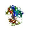
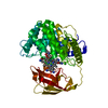
| Title | Crystallographic analysis of upper axial ligand substitutions in cobalamin bound to transcobalamin | ||||||
 Components Components | TRANSCOBALAMIN-2 | ||||||
 Keywords Keywords | TRANSPORT PROTEIN / COBALT / TRANSPORT / GLYCOPROTEIN / ION TRANSPORT / VITAMIN B12 TRANSPORT PROTEIN / COBALT TRANSPORT / BETA LIGAND SUBSTITUTION | ||||||
| Function / homology |  Function and homology information Function and homology informationTransport of RCbl within the body / cobalt ion transport / cobalamin transport / cobalamin binding / : / metal ion binding Similarity search - Function | ||||||
| Biological species |  | ||||||
| Method |  X-RAY DIFFRACTION / SYNCHROTRON / MOLECULAR REPLACEMENT / Resolution: 2.73 Å X-RAY DIFFRACTION / SYNCHROTRON / MOLECULAR REPLACEMENT / Resolution: 2.73 Å | ||||||
 Authors Authors | Wuerges, J. / Geremia, S. / Randaccio, L. | ||||||
 Citation Citation | Journal: Iubmb Life / Year: 2007 Title: Vitamin B12 Transport Proteins: Crystallographic Analysis of Beta-Axial Ligand Substitutions in Cobalamin Bound to Transcobalamin. Authors: Wuerges, J. / Geremia, S. / Fedosov, S.N. / Randaccio, L. | ||||||
| History |
| ||||||
| Remark 650 | HELIX DETERMINATION METHOD: AUTHOR PROVIDED. | ||||||
| Remark 700 | SHEET DETERMINATION METHOD: AUTHOR PROVIDED. |
- Structure visualization
| Structure viewer | Molecule: MolmilJmol/JSmol |
|---|
- Downloads & links
-Download
| PDBx/mmCIF format | 2v3n.cif.gz | 99.9 KB | Display | PDBx/mmCIF format |
|---|---|---|---|---|
| PDB format | pdb2v3n.ent.gz | 75.7 KB | Display | PDB format |
| PDBx/mmJSON format | 2v3n.json.gz | Tree view | PDBx/mmJSON format | |
| Others |  Other downloads Other downloads |
-Validation report
| Arichive directory | https://data.pdbj.org/pub/pdb/validation_reports/v3/2v3nftp://data.pdbj.org/pub/pdb/validation_reports/v3/2v3n | HTTPS FTP |
|---|
-Related structure data
| Related structure data |  2v3pC  2bbcS C: citing same article ( S: Starting model for refinement |
|---|---|
| Similar structure data |


-Links
 PDBj
PDBj


- Assembly
| Deposited unit | 
| ||||||||
|---|---|---|---|---|---|---|---|---|---|
| 1 |
| ||||||||
| Unit cell |
|
-Components
| #1: Protein | Mass: 46315.219 Da / Num. of mol.: 1 Source method: isolated from a genetically manipulated source Source: (gene. exp.)  PICHIA PASTORIS (fungus) / Strain (production host): SMD 1168 / References: UniProt: Q9XSC9 PICHIA PASTORIS (fungus) / Strain (production host): SMD 1168 / References: UniProt: Q9XSC9 | ||||
|---|---|---|---|---|---|
| #2: Chemical | ChemComp-B12 /   Mass: 1330.356 Da / Num. of mol.: 1 / Source method: obtained synthetically / Formula: C62H89CoN13O14P Mass: 1330.356 Da / Num. of mol.: 1 / Source method: obtained synthetically / Formula: C62H89CoN13O14P | ||||
| #3: Chemical | ChemComp-CYN /   Mass: 26.017 Da / Num. of mol.: 1 / Source method: obtained synthetically / Formula: CN Mass: 26.017 Da / Num. of mol.: 1 / Source method: obtained synthetically / Formula: CN | ||||
| #4: Chemical |   Mass: 35.453 Da / Num. of mol.: 3 / Source method: obtained synthetically / Formula: Cl Mass: 35.453 Da / Num. of mol.: 3 / Source method: obtained synthetically / Formula: Cl#5: Water | ChemComp-HOH / |  Mass: 18.015 Da / Num. of mol.: 174 / Source method: isolated from a natural source / Formula: H2O Mass: 18.015 Da / Num. of mol.: 174 / Source method: isolated from a natural source / Formula: H2OHas protein modification | Y | |
-Experimental details
-Experiment
| Experiment | Method: X-RAY DIFFRACTION / Number of used crystals: 1 |
|---|
- Sample preparation
| Crystal | Density Matthews: 4.1 Å3/Da / Density % sol: 69.9 % / Description: NONE |
|---|---|
| Crystal grow | pH: 8.5 Details: PROTEIN AT 0.5 MM IN 1 M NACL, 0.1 M TRIS, PH 7.5 CRYSTALLIZED FROM 28% PEG 8000, 0.2 M MAGNESIUM ACETATE, 0.1 M TRIS PH 8.5, 20% 2-METHYL-2, 4-PENTADIOL, 15 MM KCN |
-Data collection
| Diffraction | Mean temperature: 100 K |
|---|---|
| Diffraction source | Source: SYNCHROTRON / Site: ELETTRA  / Beamline: 5.2R / Wavelength: 1.2 / Beamline: 5.2R / Wavelength: 1.2 |
| Detector | Type: MARRESEARCH / Detector: CCD / Date: Apr 4, 2007 |
| Radiation | Monochromator: DOUBLE CRYSTAL SI(220) / Protocol: SINGLE WAVELENGTH / Monochromatic (M) / Laue (L): M / Scattering type: x-ray |
| Radiation wavelength | Wavelength: 1.2 Å / Relative weight: 1 |
| Reflection | Resolution: 2.7→20.7 Å / Num. obs: 21370 / % possible obs: 99.3 % / Observed criterion σ(I): 0 / Redundancy: 3.1 % / Rmerge(I) obs: 0.12 / Net I/σ(I): 9.7 |
| Reflection shell | Resolution: 2.7→2.85 Å / Redundancy: 3.1 % / Rmerge(I) obs: 0.75 / Mean I/σ(I) obs: 1.7 / % possible all: 99.9 |
- Processing
| Software |
| ||||||||||||||||||||||||||||||||||||||||||||||||||||||||||||||||||||||||||||||||||||||||||||||||||||||||||||||||||||||||||||||||||||||||||||||||||||||||||||||||||||||||||||||||||||||
|---|---|---|---|---|---|---|---|---|---|---|---|---|---|---|---|---|---|---|---|---|---|---|---|---|---|---|---|---|---|---|---|---|---|---|---|---|---|---|---|---|---|---|---|---|---|---|---|---|---|---|---|---|---|---|---|---|---|---|---|---|---|---|---|---|---|---|---|---|---|---|---|---|---|---|---|---|---|---|---|---|---|---|---|---|---|---|---|---|---|---|---|---|---|---|---|---|---|---|---|---|---|---|---|---|---|---|---|---|---|---|---|---|---|---|---|---|---|---|---|---|---|---|---|---|---|---|---|---|---|---|---|---|---|---|---|---|---|---|---|---|---|---|---|---|---|---|---|---|---|---|---|---|---|---|---|---|---|---|---|---|---|---|---|---|---|---|---|---|---|---|---|---|---|---|---|---|---|---|---|---|---|---|---|
| Refinement | Method to determine structure: MOLECULAR REPLACEMENT Starting model: PDB ENTRY 2BBC Resolution: 2.73→20.7 Å / Cor.coef. Fo:Fc: 0.928 / Cor.coef. Fo:Fc free: 0.898 / SU B: 19.725 / SU ML: 0.212 / TLS residual ADP flag: LIKELY RESIDUAL / Cross valid method: THROUGHOUT / σ(F): 0 / ESU R: 0.448 / ESU R Free: 0.291 / Stereochemistry target values: MAXIMUM LIKELIHOOD Details: HYDROGENS HAVE BEEN ADDED IN THE RIDING POSITIONS. AMINO ACIDS FROM LYS168 TO VAL176 WERE NOT MODELLED DUE TO DISORDER. HIS175 AS UPPER AXIAL LIGAND OF COBALAMIN HAS BEEN DISPLACED BY ...Details: HYDROGENS HAVE BEEN ADDED IN THE RIDING POSITIONS. AMINO ACIDS FROM LYS168 TO VAL176 WERE NOT MODELLED DUE TO DISORDER. HIS175 AS UPPER AXIAL LIGAND OF COBALAMIN HAS BEEN DISPLACED BY EXTERNALLY SUPPLIED CYANIDE.
| ||||||||||||||||||||||||||||||||||||||||||||||||||||||||||||||||||||||||||||||||||||||||||||||||||||||||||||||||||||||||||||||||||||||||||||||||||||||||||||||||||||||||||||||||||||||
| Solvent computation | Ion probe radii: 0.8 Å / Shrinkage radii: 0.8 Å / VDW probe radii: 1.4 Å / Solvent model: MASK BULK SOLVENT | ||||||||||||||||||||||||||||||||||||||||||||||||||||||||||||||||||||||||||||||||||||||||||||||||||||||||||||||||||||||||||||||||||||||||||||||||||||||||||||||||||||||||||||||||||||||
| Displacement parameters | Biso mean: 43.4 Å2
| ||||||||||||||||||||||||||||||||||||||||||||||||||||||||||||||||||||||||||||||||||||||||||||||||||||||||||||||||||||||||||||||||||||||||||||||||||||||||||||||||||||||||||||||||||||||
| Refinement step | Cycle: LAST / Resolution: 2.73→20.7 Å
| ||||||||||||||||||||||||||||||||||||||||||||||||||||||||||||||||||||||||||||||||||||||||||||||||||||||||||||||||||||||||||||||||||||||||||||||||||||||||||||||||||||||||||||||||||||||
| Refine LS restraints |
|