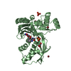
[English] 日本語
Yorodumi
- PDB-2v2z: IspE in complex with ADP and CDPME -

+
Open data


ID or keywords:

Loading...

-
Basic information

Entry
Database: PDB / ID: 2v2z
TitleIspE in complex with ADP and CDPME
Components4-DIPHOSPHOCYTIDYL- ...
KeywordsTRANSFERASE / 4-DIPHOSPHOCYTIDYL-2C-METHYL-D-ERYTHRITOL / AQUIFEX AEOLICUS / NUCLEOTIDE-BINDING / ISOPRENE BIOSYNTHESIS / KINASE / ATP-BINDING / NON-MEVALONATE
Function / homology
Function and homology information


4-(cytidine 5'-diphospho)-2-C-methyl-D-erythritol kinase / 4-(cytidine 5'-diphospho)-2-C-methyl-D-erythritol kinase activity / isopentenyl diphosphate biosynthetic process, methylerythritol 4-phosphate pathway / terpenoid biosynthetic process / phosphorylation / ATP binding
Similarity search - Function
4-diphosphocytidyl-2C-methyl-D-erythritol kinase / GHMP kinase, C-terminal domain / GHMP kinase N-terminal domain / GHMP kinases N terminal domain / GHMP kinase, C-terminal domain superfamily / Ribosomal Protein S5; domain 2 - #10 / Ribosomal Protein S5; domain 2 / Ribosomal protein S5 domain 2-type fold, subgroup / Ribosomal protein S5 domain 2-type fold / Alpha-Beta Plaits ...4-diphosphocytidyl-2C-methyl-D-erythritol kinase / GHMP kinase, C-terminal domain / GHMP kinase N-terminal domain / GHMP kinases N terminal domain / GHMP kinase, C-terminal domain superfamily / Ribosomal Protein S5; domain 2 - #10 / Ribosomal Protein S5; domain 2 / Ribosomal protein S5 domain 2-type fold, subgroup / Ribosomal protein S5 domain 2-type fold / Alpha-Beta Plaits / 2-Layer Sandwich / Alpha Beta
Similarity search - Domain/homology
ADENOSINE-5'-DIPHOSPHATE / 4-DIPHOSPHOCYTIDYL-2-C-METHYL-D-ERYTHRITOL / 4-diphosphocytidyl-2-C-methyl-D-erythritol kinase
Similarity search - Component
Biological speciesAQUIFEX AEOLICUS (bacteria)
MethodX-RAY DIFFRACTION / SYNCHROTRON / MOLECULAR REPLACEMENT / Resolution: 2.25 Å
AuthorsSgraja, T. / Alphey, M.S. / Hunter, W.N.
CitationJournal: FEBS J. / Year: 2008
Title: Characterization of Aquifex Aeolicus 4-Diphosphocytidyl-2C-Methyl-D-Erythritol Kinase -Ligand Recognition in a Template for Antimicrobial Drug Discovery.
Authors: Sgraja, T. / Alphey, M.S. / Ghilagaber, S. / Marquez, R. / Robertson, M.N. / Hemmings, J.L. / Lauw, S. / Rohdich, F. / Bacher, A. / Eisenreich, W. / Illarionova, V. / Hunter, W.N.
History
DepositionJun 8, 2007Deposition site: PDBE / Processing site: PDBE
Revision 1.0Jun 19, 2007Provider: repository / Type: Initial release
Revision 1.1May 8, 2011Group: Version format compliance
Revision 1.2Jul 13, 2011Group: Version format compliance
Remark 700 SHEET THE SHEET STRUCTURE OF THIS MOLECULE IS BIFURCATED. IN ORDER TO REPRESENT THIS FEATURE IN ... SHEET THE SHEET STRUCTURE OF THIS MOLECULE IS BIFURCATED. IN ORDER TO REPRESENT THIS FEATURE IN THE SHEET RECORDS BELOW, TWO SHEETS ARE DEFINED.

-
Structure visualization

Structure viewerMolecule:
MolmilJmol/JSmol

Downloads & links

-
Assembly

Deposited unit
A: 4-DIPHOSPHOCYTIDYL-2C-METHYL-D-ERYTHRITOL KINASE
B: 4-DIPHOSPHOCYTIDYL-2C-METHYL-D-ERYTHRITOL KINASE
hetero molecules


Theoretical massNumber of molelcules
Total (without water)62,49212
Polymers60,2012
Non-polymers2,29210
Water5,513306
1
A: 4-DIPHOSPHOCYTIDYL-2C-METHYL-D-ERYTHRITOL KINASE
hetero molecules


Theoretical massNumber of molelcules
Total (without water)31,2766
Polymers30,1001
Non-polymers1,1765
Water181
TypeNameSymmetry operationNumber
identity operation1_555x,y,z1
MethodPQS
2
B: 4-DIPHOSPHOCYTIDYL-2C-METHYL-D-ERYTHRITOL KINASE
hetero molecules


Theoretical massNumber of molelcules
Total (without water)31,2166
Polymers30,1001
Non-polymers1,1155
Water181
TypeNameSymmetry operationNumber
identity operation1_555x,y,z1
MethodPQS
Unit cell
Length a, b, c (Å)136.994, 136.994, 136.994
Angle α, β, γ (deg.)90.00, 90.00, 90.00
Int Tables number198
Space group name H-MP213
Components on special symmetry positions
IDModelComponents
11B-2010-

HOH

21B-2024-

HOH

-
Components

-
4-DIPHOSPHOCYTIDYL- ... , 1 types, 2 molecules AB

#1: Protein 4-DIPHOSPHOCYTIDYL-2C-METHYL-D-ERYTHRITOL KINASE / CMK / 4-CYTIDINE-5'-DIPHOSPHO-2-C-METHYL-D-ERYTHRITOL KINASE


Mass: 30100.377 Da / Num. of mol.: 2
Source method: isolated from a genetically manipulated source
Source: (gene. exp.) AQUIFEX AEOLICUS (bacteria) / Plasmid: PET15B / Production host: ESCHERICHIA COLI (E. coli) / Strain (production host): BL21(DE3)
References: UniProt: O67060, 4-(cytidine 5'-diphospho)-2-C-methyl-D-erythritol kinase

-
Non-polymers , 5 types, 316 molecules

#2: Chemical ChemComp-CDM / 4-DIPHOSPHOCYTIDYL-2-C-METHYL-D-ERYTHRITOL / 4-Diphosphocytidyl-2-C-methylerythritol


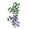
Mass: 521.308 Da / Num. of mol.: 2 / Source method: obtained synthetically / Formula: C14H25N3O14P2
#3: Chemical ChemComp-ADP / ADENOSINE-5'-DIPHOSPHATE / Adenosine diphosphate


Mass: 427.201 Da / Num. of mol.: 2 / Source method: obtained synthetically / Formula: C10H15N5O10P2 / Comment: ADP, energy-carrying molecule*YM
#4: Chemical ChemComp-SO4 / SULFATE ION / Sulfate


Mass: 96.063 Da / Num. of mol.: 3 / Source method: obtained synthetically / Formula: SO4
#5: Chemical ChemComp-CL / CHLORIDE ION / Chloride


Mass: 35.453 Da / Num. of mol.: 3 / Source method: obtained synthetically / Formula: Cl
#6: Water ChemComp-HOH / water / Water


Mass: 18.015 Da / Num. of mol.: 306 / Source method: isolated from a natural source / Formula: H2O

-
Details

Sequence detailsRESIDUES -2 TO 0 ARE RESIDUAL FROM HIS-TAG REMOVAL

-
Experimental details

-
Experiment

ExperimentMethod: X-RAY DIFFRACTION / Number of used crystals: 1

-
Sample preparation

CrystalDensity Matthews: 3.6 Å3/Da / Density % sol: 65.82 %
Crystal growpH: 6.5 / Details: 0.1 M MES PH 6.5, 0.2 M NACL AND 1.5 M (NH4)2SO4

-
Data collection

DiffractionMean temperature: 100 K
Diffraction sourceSource: SYNCHROTRON / Site: ESRF / Beamline: ID23-1 / Wavelength: 0.873
DetectorType: ADSC CCD / Detector: CCD / Details: TOROID MIRROR
RadiationMonochromator: SI111 / Protocol: SINGLE WAVELENGTH / Monochromatic (M) / Laue (L): M / Scattering type: x-ray
Radiation wavelengthWavelength: 0.873 Å / Relative weight: 1
ReflectionResolution: 2.25→40 Å / Num. obs: 40884 / % possible obs: 100 % / Redundancy: 7.4 % / Rmerge(I) obs: 0.13 / Net I/σ(I): 12.1
Reflection shellHighest resolution: 2.25 Å / Redundancy: 7.6 % / Rmerge(I) obs: 0.49 / Mean I/σ(I) obs: 2.9 / % possible all: 100

-
Processing

Software
NameVersionClassification
REFMAC5.2.0019refinement
MOSFLMdata reduction
SCALAdata scaling
MOLREPphasing
RefinementMethod to determine structure: MOLECULAR REPLACEMENT / Resolution: 2.25→39.56 Å / Cor.coef. Fo:Fc: 0.936 / Cor.coef. Fo:Fc free: 0.927 / SU B: 5.123 / SU ML: 0.131 / Cross valid method: THROUGHOUT / ESU R: 0.226 / ESU R Free: 0.183 / Stereochemistry target values: MAXIMUM LIKELIHOOD / Details: HYDROGENS HAVE BEEN ADDED IN THE RIDING POSITIONS.
RfactorNum. reflection% reflectionSelection details
Rfree0.23 2047 5 %RANDOM
Rwork0.202 ---
obs0.204 38809 100 %-
Solvent computationIon probe radii: 0.8 Å / Shrinkage radii: 0.8 Å / VDW probe radii: 1.4 Å / Solvent model: MASK
Displacement parametersBiso mean: 31.38 Å2
Refinement stepCycle: LAST / Resolution: 2.25→39.56 Å
ProteinNucleic acidLigandSolventTotal
Num. atoms4237 0 138 306 4681
Refine LS restraints
Refine-IDTypeDev idealDev ideal targetNumber
X-RAY DIFFRACTIONr_bond_refined_d0.0080.0224508
X-RAY DIFFRACTIONr_bond_other_d
X-RAY DIFFRACTIONr_angle_refined_deg1.1682.0276128
X-RAY DIFFRACTIONr_angle_other_deg
X-RAY DIFFRACTIONr_dihedral_angle_1_deg5.5715549
X-RAY DIFFRACTIONr_dihedral_angle_2_deg34.31924.359195
X-RAY DIFFRACTIONr_dihedral_angle_3_deg14.80715788
X-RAY DIFFRACTIONr_dihedral_angle_4_deg21.4991527
X-RAY DIFFRACTIONr_chiral_restr0.0660.2679
X-RAY DIFFRACTIONr_gen_planes_refined0.0030.023329
X-RAY DIFFRACTIONr_gen_planes_other
X-RAY DIFFRACTIONr_nbd_refined0.1760.21923
X-RAY DIFFRACTIONr_nbd_other
X-RAY DIFFRACTIONr_nbtor_refined0.30.23046
X-RAY DIFFRACTIONr_nbtor_other
X-RAY DIFFRACTIONr_xyhbond_nbd_refined0.1250.2297
X-RAY DIFFRACTIONr_xyhbond_nbd_other
X-RAY DIFFRACTIONr_metal_ion_refined
X-RAY DIFFRACTIONr_metal_ion_other
X-RAY DIFFRACTIONr_symmetry_vdw_refined0.170.274
X-RAY DIFFRACTIONr_symmetry_vdw_other
X-RAY DIFFRACTIONr_symmetry_hbond_refined0.1850.214
X-RAY DIFFRACTIONr_symmetry_hbond_other
X-RAY DIFFRACTIONr_symmetry_metal_ion_refined
X-RAY DIFFRACTIONr_symmetry_metal_ion_other
X-RAY DIFFRACTIONr_mcbond_it0.4771.52787
X-RAY DIFFRACTIONr_mcbond_other
X-RAY DIFFRACTIONr_mcangle_it0.84724346
X-RAY DIFFRACTIONr_mcangle_other
X-RAY DIFFRACTIONr_scbond_it1.11531978
X-RAY DIFFRACTIONr_scbond_other
X-RAY DIFFRACTIONr_scangle_it1.8734.51776
X-RAY DIFFRACTIONr_scangle_other
X-RAY DIFFRACTIONr_long_range_B_refined
X-RAY DIFFRACTIONr_long_range_B_other
X-RAY DIFFRACTIONr_rigid_bond_restr
X-RAY DIFFRACTIONr_sphericity_free
X-RAY DIFFRACTIONr_sphericity_bonded
LS refinement shellResolution: 2.25→2.31 Å / Total num. of bins used: 20 /
RfactorNum. reflection
Rfree0.296 159
Rwork0.248 2836

+
About Yorodumi

-
News

-
Feb 9, 2022. New format data for meta-information of EMDB entries

New format data for meta-information of EMDB entries

  • Version 3 of the EMDB header file is now the official format.
  • The previous official version 1.9 will be removed from the archive.

Related info.:EMDB header

External links:wwPDB to switch to version 3 of the EMDB data model

-
Aug 12, 2020. Covid-19 info

Covid-19 info

URL: https://pdbj.org/emnavi/covid19.php

New page: Covid-19 featured information page in EM Navigator.

Related info.:Covid-19 info / Mar 5, 2020. Novel coronavirus structure data

+
Mar 5, 2020. Novel coronavirus structure data

Novel coronavirus structure data

Related info.:Yorodumi Speices / Aug 12, 2020. Covid-19 info

External links:COVID-19 featured content - PDBj / Molecule of the Month (242):Coronavirus Proteases

+
Jan 31, 2019. EMDB accession codes are about to change! (news from PDBe EMDB page)

EMDB accession codes are about to change! (news from PDBe EMDB page)

  • The allocation of 4 digits for EMDB accession codes will soon come to an end. Whilst these codes will remain in use, new EMDB accession codes will include an additional digit and will expand incrementally as the available range of codes is exhausted. The current 4-digit format prefixed with “EMD-” (i.e. EMD-XXXX) will advance to a 5-digit format (i.e. EMD-XXXXX), and so on. It is currently estimated that the 4-digit codes will be depleted around Spring 2019, at which point the 5-digit format will come into force.
  • The EM Navigator/Yorodumi systems omit the EMD- prefix.

Related info.:Q: What is EMD? / ID/Accession-code notation in Yorodumi/EM Navigator

External links:EMDB Accession Codes are Changing Soon! / Contact to PDBj

+
Jul 12, 2017. Major update of PDB

Major update of PDB

  • wwPDB released updated PDB data conforming to the new PDBx/mmCIF dictionary.
  • This is a major update changing the version number from 4 to 5, and with Remediation, in which all the entries are updated.
  • In this update, many items about electron microscopy experimental information are reorganized (e.g. em_software).
  • Now, EM Navigator and Yorodumi are based on the updated data.

External links:wwPDB Remediation / Enriched Model Files Conforming to OneDep Data Standards Now Available in the PDB FTP Archive

-
Yorodumi

Thousand views of thousand structures

  • Yorodumi is a browser for structure data from EMDB, PDB, SASBDB, etc.
  • This page is also the successor to EM Navigator detail page, and also detail information page/front-end page for Omokage search.
  • The word "yorodu" (or yorozu) is an old Japanese word meaning "ten thousand". "mi" (miru) is to see.

Related info.:EMDB / PDB / SASBDB / Comparison of 3 databanks / Yorodumi Search / Aug 31, 2016. New EM Navigator & Yorodumi / Yorodumi Papers / Jmol/JSmol / Function and homology information / Changes in new EM Navigator and Yorodumi

Read more