 Movie
Movie Controller

[English] 日本語
 Yorodumi
- PDB-2v1l: Structure of the conserved hypothetical protein VC1805 from patho... -
+ Open data

- Basic information
| Entry | Database: PDB / ID: 2v1l | ||||||
|---|---|---|---|---|---|---|---|
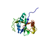
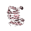
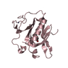
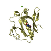
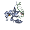
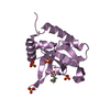
| Title | Structure of the conserved hypothetical protein VC1805 from pathogenicity island VPI-2 of Vibrio cholerae O1 biovar eltor str. N16961 shares structural homology with the human P32 protein | ||||||
 Components Components | HYPOTHETICAL PROTEIN | ||||||
 Keywords Keywords | UNKNOWN FUNCTION / PATHOGENICITY ISLAND / HYPOTHETICAL PROTEIN | ||||||
| Function / homology | Protein of unknown function DUF2787 / Protein of unknown function DUF2787 / Protein of unknown function (DUF2787) / Nuclear Transport Factor 2; Chain: A, / Roll / Alpha Beta / DUF2787 domain-containing protein Function and homology information Function and homology information | ||||||
| Biological species |   VIBRIO CHOLERAE (bacteria) VIBRIO CHOLERAE (bacteria) | ||||||
| Method |  X-RAY DIFFRACTION / SYNCHROTRON / MIRAS / Resolution: 2.13 Å X-RAY DIFFRACTION / SYNCHROTRON / MIRAS / Resolution: 2.13 Å | ||||||
 Authors Authors | Sheikh, M.A. / Potter, J.A. / Johnson, K.A. / Boyd, E.F. / Taylor, G.L. | ||||||
 Citation Citation | Journal: Proteins / Year: 2008 Title: Crystal Structure of Vc1805, a Conserved Hypothetical Protein from a Vibrio Cholerae Pathogenicity Island, Reveals Homology to Human P32. Authors: Sheikh, M.A. / Potter, J.A. / Johnson, K.A. / Sim, R.B. / Boyd, E.F. / Taylor, G.L. | ||||||
| History |
|
- Structure visualization
| Structure viewer | Molecule: MolmilJmol/JSmol |
|---|
- Downloads & links
-Download
| PDBx/mmCIF format | 2v1l.cif.gz | 38.5 KB | Display | PDBx/mmCIF format |
|---|---|---|---|---|
| PDB format | pdb2v1l.ent.gz | 27 KB | Display | PDB format |
| PDBx/mmJSON format | 2v1l.json.gz | Tree view | PDBx/mmJSON format | |
| Others |  Other downloads Other downloads |
-Validation report
| Summary document | 2v1l_validation.pdf.gz | 427.8 KB | Display | wwPDB validaton report |
|---|---|---|---|---|
| Full document | 2v1l_full_validation.pdf.gz | 430.3 KB | Display | |
| Data in XML | 2v1l_validation.xml.gz | 7.7 KB | Display | |
| Data in CIF | 2v1l_validation.cif.gz | 9.6 KB | Display | |
| Arichive directory | https://data.pdbj.org/pub/pdb/validation_reports/v1/2v1lftp://data.pdbj.org/pub/pdb/validation_reports/v1/2v1l | HTTPS FTP |
-Related structure data
| Similar structure data |
|---|


-Links
 PDBj
PDBj- Assembly
| Deposited unit | 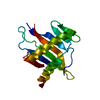
| ||||||||
|---|---|---|---|---|---|---|---|---|---|
| 1 |
| ||||||||
| Unit cell |
|
-Components
| #1: Protein | Mass: 16846.613 Da / Num. of mol.: 1 Source method: isolated from a genetically manipulated source Source: (gene. exp.) VIBRIO CHOLERAE (bacteria) / Strain: O1 / Variant: BIOVAR ELTOR STR. N16961 / Production host: |
|---|---|
| #2: Water | ChemComp-HOH /  Mass: 18.015 Da / Num. of mol.: 35 / Source method: isolated from a natural source / Formula: H2O Mass: 18.015 Da / Num. of mol.: 35 / Source method: isolated from a natural source / Formula: H2O |
-Experimental details
-Experiment
| Experiment | Method: X-RAY DIFFRACTION |
|---|
- Sample preparation
| Crystal | Density Matthews: 2.46 Å3/Da / Density % sol: 44 % / Description: NONE |
|---|---|
| Crystal grow | pH: 7.5 / Details: pH 7.5 |
-Data collection
| Diffraction | Mean temperature: 100 K |
|---|---|
| Diffraction source | Source: SYNCHROTRON / Site: ESRF  / Beamline: ID14-1 / Wavelength: 0.934 / Beamline: ID14-1 / Wavelength: 0.934 |
| Detector | Type: ADSC CCD / Detector: CCD |
| Radiation | Protocol: SINGLE WAVELENGTH / Monochromatic (M) / Laue (L): M / Scattering type: x-ray |
| Radiation wavelength | Wavelength: 0.934 Å / Relative weight: 1 |
| Reflection | Resolution: 2.13→39.16 Å / Num. obs: 8131 / % possible obs: 99 % / Observed criterion σ(I): 2 / Redundancy: 8.8 % / Rmerge(I) obs: 0.06 / Net I/σ(I): 23 |
| Reflection shell | Resolution: 2.13→2.25 Å / Redundancy: 9 % / Rmerge(I) obs: 0.38 / Mean I/σ(I) obs: 6.4 / % possible all: 99 |
- Processing
| Software |
| ||||||||||||||||||||||||||||||||||||||||||||||||||||||||||||||||||||||||||||||||||||||||||||||||||||||||||||||||||||||||||||||||||||||||||||||||||||||||||||||||||||||||||||||||||||||
|---|---|---|---|---|---|---|---|---|---|---|---|---|---|---|---|---|---|---|---|---|---|---|---|---|---|---|---|---|---|---|---|---|---|---|---|---|---|---|---|---|---|---|---|---|---|---|---|---|---|---|---|---|---|---|---|---|---|---|---|---|---|---|---|---|---|---|---|---|---|---|---|---|---|---|---|---|---|---|---|---|---|---|---|---|---|---|---|---|---|---|---|---|---|---|---|---|---|---|---|---|---|---|---|---|---|---|---|---|---|---|---|---|---|---|---|---|---|---|---|---|---|---|---|---|---|---|---|---|---|---|---|---|---|---|---|---|---|---|---|---|---|---|---|---|---|---|---|---|---|---|---|---|---|---|---|---|---|---|---|---|---|---|---|---|---|---|---|---|---|---|---|---|---|---|---|---|---|---|---|---|---|---|---|
| Refinement | Method to determine structure: MIRAS Starting model: NONE Resolution: 2.13→39.16 Å / Cor.coef. Fo:Fc: 0.924 / Cor.coef. Fo:Fc free: 0.871 / SU B: 6.663 / SU ML: 0.18 / Cross valid method: THROUGHOUT / ESU R: 0.274 / ESU R Free: 0.251 / Stereochemistry target values: MAXIMUM LIKELIHOOD / Details: HYDROGENS HAVE BEEN ADDED IN THE RIDING POSITIONS.
| ||||||||||||||||||||||||||||||||||||||||||||||||||||||||||||||||||||||||||||||||||||||||||||||||||||||||||||||||||||||||||||||||||||||||||||||||||||||||||||||||||||||||||||||||||||||
| Solvent computation | Ion probe radii: 0.8 Å / Shrinkage radii: 0.8 Å / VDW probe radii: 1.2 Å / Solvent model: MASK | ||||||||||||||||||||||||||||||||||||||||||||||||||||||||||||||||||||||||||||||||||||||||||||||||||||||||||||||||||||||||||||||||||||||||||||||||||||||||||||||||||||||||||||||||||||||
| Displacement parameters | Biso mean: 39.21 Å2
| ||||||||||||||||||||||||||||||||||||||||||||||||||||||||||||||||||||||||||||||||||||||||||||||||||||||||||||||||||||||||||||||||||||||||||||||||||||||||||||||||||||||||||||||||||||||
| Refinement step | Cycle: LAST / Resolution: 2.13→39.16 Å
| ||||||||||||||||||||||||||||||||||||||||||||||||||||||||||||||||||||||||||||||||||||||||||||||||||||||||||||||||||||||||||||||||||||||||||||||||||||||||||||||||||||||||||||||||||||||
| Refine LS restraints |
|