 ムービー
ムービー コントローラー


+ データを開く

- 基本情報
基本情報
| 登録情報 | データベース: PDB / ID: 2rgz | ||||||
|---|---|---|---|---|---|---|---|
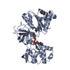
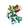
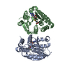
| タイトル | Ensemble refinement of the protein crystal structure of human heme oxygenase-2 C127A (HO-2) with bound heme | ||||||
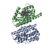
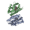
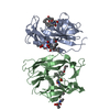
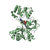
 要素 要素 | Heme oxygenase 2 | ||||||
 キーワード キーワード | OXIDOREDUCTASE / Ensemble Refinement / Refinement Methodology Development / Structural Genomics Medical Relevance / Structural Genomics Community Request / HO-2 / HEME Oxygenase / Endoplasmic reticulum / Iron / Metal-binding / Microsome / Structural Genomics / Protein Structure Initiative / PSI / Center for Eukaryotic Structural Genomics / CESG | ||||||
| 機能・相同性 |  機能・相同性情報 機能・相同性情報heme oxygenase (biliverdin-producing) / heme oxidation / heme oxygenase (decyclizing) activity / heme catabolic process / Heme degradation / RHOA GTPase cycle / specific granule membrane / Iron uptake and transport / Cytoprotection by HMOX1 / response to oxidative stress ...heme oxygenase (biliverdin-producing) / heme oxidation / heme oxygenase (decyclizing) activity / heme catabolic process / Heme degradation / RHOA GTPase cycle / specific granule membrane / Iron uptake and transport / Cytoprotection by HMOX1 / response to oxidative stress / response to hypoxia / heme binding / Neutrophil degranulation / endoplasmic reticulum membrane / membrane / metal ion binding / plasma membrane 類似検索 - 分子機能 | ||||||
| 生物種 |  Homo sapiens (ヒト) Homo sapiens (ヒト) | ||||||
| 手法 |  X線回折 / シンクロトロン / Multi-conformer re-refinement / 解像度: 2.61 Å X線回折 / シンクロトロン / Multi-conformer re-refinement / 解像度: 2.61 Å | ||||||
 データ登録者 データ登録者 | Bianchetti, C.M. / Bingman, C.A. / Bitto, E. / Wesenberg, G.E. / Phillips Jr., G.N. / Center for Eukaryotic Structural Genomics (CESG) | ||||||
 引用 引用 | ジャーナル: J.Biol.Chem. / 年: 2007 タイトル: Comparison of Apo- and Heme-bound Crystal Structures of a Truncated Human Heme Oxygenase-2. 著者: Bianchetti, C.M. / Yi, L. / Ragsdale, S.W. / Phillips Jr., G.N. | ||||||
| 履歴 |
|
- 構造の表示
構造の表示
| 構造ビューア | 分子: MolmilJmol/JSmol |
|---|
- ダウンロードとリンク
-ダウンロード
| PDBx/mmCIF形式 | 2rgz.cif.gz | 1.3 MB | 表示 | PDBx/mmCIF形式 |
|---|---|---|---|---|
| PDB形式 | pdb2rgz.ent.gz | 1.1 MB | 表示 | PDB形式 |
| PDBx/mmJSON形式 | 2rgz.json.gz | ツリー表示 | PDBx/mmJSON形式 | |
| その他 |  その他のダウンロード その他のダウンロード |
-検証レポート
| 文書・要旨 | 2rgz_validation.pdf.gz | 1 MB | 表示 | wwPDB検証レポート |
|---|---|---|---|---|
| 文書・詳細版 | 2rgz_full_validation.pdf.gz | 1.1 MB | 表示 | |
| XML形式データ | 2rgz_validation.xml.gz | 131.6 KB | 表示 | |
| CIF形式データ | 2rgz_validation.cif.gz | 172.4 KB | 表示 | |
| アーカイブディレクトリ | https://data.pdbj.org/pub/pdb/validation_reports/rg/2rgzftp://data.pdbj.org/pub/pdb/validation_reports/rg/2rgz | HTTPS FTP |
-関連構造データ


-リンク
 PDBj
PDBj


- 集合体
集合体
| 登録構造単位 | 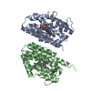
| ||||||||
|---|---|---|---|---|---|---|---|---|---|
| 1 |
| ||||||||
| 2 | 
| ||||||||
| 3 | 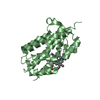
| ||||||||
| 単位格子 |
| ||||||||
| モデル数 | 16 |
-要素
| #1: タンパク質 | 分子量: 30491.271 Da / 分子数: 2 / 変異: C127A / 由来タイプ: 組換発現 / 由来: (組換発現) Homo sapiens (ヒト) / 遺伝子: HMOX2, HO2 / プラスミド: pGEX 4T-2 / 生物種 (発現宿主): Escherichia coli / 発現宿主:  参照: UniProt: P30519, heme oxygenase (biliverdin-producing) #2: 化合物 |   分子量: 616.487 Da / 分子数: 2 / 由来タイプ: 合成 / 式: C34H32FeN4O4 分子量: 616.487 Da / 分子数: 2 / 由来タイプ: 合成 / 式: C34H32FeN4O4#3: 水 | ChemComp-HOH / |  分子量: 18.015 Da / 分子数: 83 / 由来タイプ: 天然 / 式: H2O 分子量: 18.015 Da / 分子数: 83 / 由来タイプ: 天然 / 式: H2O |
|---|
-実験情報
-実験
| 実験 | 手法: X線回折 / 使用した結晶の数: 1 |
|---|
- 試料調製
試料調製
| 結晶 | マシュー密度: 2.56 Å3/Da / 溶媒含有率: 51.94 % |
|---|---|
| 結晶化 | 温度: 277 K / 手法: 蒸気拡散法, ハンギングドロップ法 詳細: Protein solution (5 mg/ml Protein, 1:1 ratio Heme, 2% DMSO, 0.050 M Potassium chloride, 0.050 M Tris-HCl pH 7.5) mixed in a 1.5:1 ratio with the Well solution (33% PEG DME 500, 0.020 M ...詳細: Protein solution (5 mg/ml Protein, 1:1 ratio Heme, 2% DMSO, 0.050 M Potassium chloride, 0.050 M Tris-HCl pH 7.5) mixed in a 1.5:1 ratio with the Well solution (33% PEG DME 500, 0.020 M Magnesium chloride, 0.10 M HEPES pH 7.5). Cryoprotected with well solution, VAPOR DIFFUSION, HANGING DROP, temperature 277K |
-データ収集
| 回折 | 平均測定温度: 100 K | |||||||||||||||||||||||||||||||||||||||||||||||||||||||||||||||||||||||||||||
|---|---|---|---|---|---|---|---|---|---|---|---|---|---|---|---|---|---|---|---|---|---|---|---|---|---|---|---|---|---|---|---|---|---|---|---|---|---|---|---|---|---|---|---|---|---|---|---|---|---|---|---|---|---|---|---|---|---|---|---|---|---|---|---|---|---|---|---|---|---|---|---|---|---|---|---|---|---|---|
| 放射光源 | 由来: シンクロトロン / サイト: APS  / ビームライン: 23-ID-B / 波長: 0.97946 Å / ビームライン: 23-ID-B / 波長: 0.97946 Å | |||||||||||||||||||||||||||||||||||||||||||||||||||||||||||||||||||||||||||||
| 検出器 | タイプ: MARMOSAIC 300 mm CCD / 検出器: CCD / 日付: 2007年7月6日 / 詳細: Adjustable focus K-B pair Si plus Pt, Rh coatings | |||||||||||||||||||||||||||||||||||||||||||||||||||||||||||||||||||||||||||||
| 放射 | モノクロメーター: Double crystal cryo-cooled Si(111) プロトコル: SINGLE WAVELENGTH / 単色(M)・ラウエ(L): M / 散乱光タイプ: x-ray | |||||||||||||||||||||||||||||||||||||||||||||||||||||||||||||||||||||||||||||
| 放射波長 | 波長: 0.97946 Å / 相対比: 1 | |||||||||||||||||||||||||||||||||||||||||||||||||||||||||||||||||||||||||||||
| 反射 | 解像度: 2.32→42.413 Å / Num. obs: 24030 / % possible obs: 85.6 % / 冗長度: 11.6 % / Rmerge(I) obs: 0.083 / Χ2: 1.039 / Net I/σ(I): 16.268 | |||||||||||||||||||||||||||||||||||||||||||||||||||||||||||||||||||||||||||||
| 反射 シェル | Diffraction-ID: 1
|
- 解析
解析
| ソフトウェア |
| ||||||||||||||||||||||||||||||||||||||||||||||||||||||||||||||||||||||
|---|---|---|---|---|---|---|---|---|---|---|---|---|---|---|---|---|---|---|---|---|---|---|---|---|---|---|---|---|---|---|---|---|---|---|---|---|---|---|---|---|---|---|---|---|---|---|---|---|---|---|---|---|---|---|---|---|---|---|---|---|---|---|---|---|---|---|---|---|---|---|---|
| 精密化 | 構造決定の手法: Multi-conformer re-refinement 開始モデル: PDB entry 2QPP 解像度: 2.61→39.02 Å / Rfactor Rfree error: 0.008 / Data cutoff high absF: 1772722.5 / Data cutoff low absF: 0 / Isotropic thermal model: RESTRAINED / 交差検証法: THROUGHOUT / σ(F): 0 立体化学のターゲット値: maximum likelihood using amplitudes 詳細: This PDB entry is a re-refinement using an ensemble model of the previously deposited single-conformer structure 2qpp and the first data set in the deposited structure factor file for 2qpp ...詳細: This PDB entry is a re-refinement using an ensemble model of the previously deposited single-conformer structure 2qpp and the first data set in the deposited structure factor file for 2qpp along with the R-free set defined therein. The coordinates were generated by an automated protocol from an initial model consisting of 16 identical copies of the protein and non-water hetero-atoms assigned fractional occupancies adding up to one, and a single copy of the solvent molecules. Refinement was carried out with all the conformers present simultaneously and with the potential energy terms corresponding to interactions between the different conformers excluded. The helix and sheet records were calculated using coordinates from the first conformer only. The structure visualization program PYMOL is well-suited for directly viewing the ensemble model presented in this PDB file.
| ||||||||||||||||||||||||||||||||||||||||||||||||||||||||||||||||||||||
| 溶媒の処理 | 溶媒モデル: FLAT MODEL / Bsol: 53.313 Å2 / ksol: 0.329 e/Å3 | ||||||||||||||||||||||||||||||||||||||||||||||||||||||||||||||||||||||
| 原子変位パラメータ | Biso mean: 56.5 Å2
| ||||||||||||||||||||||||||||||||||||||||||||||||||||||||||||||||||||||
| Refine analyze |
| ||||||||||||||||||||||||||||||||||||||||||||||||||||||||||||||||||||||
| 精密化ステップ | サイクル: LAST / 解像度: 2.61→39.02 Å
| ||||||||||||||||||||||||||||||||||||||||||||||||||||||||||||||||||||||
| 拘束条件 |
| ||||||||||||||||||||||||||||||||||||||||||||||||||||||||||||||||||||||
| LS精密化 シェル | Refine-ID: X-RAY DIFFRACTION / Total num. of bins used: 6
| ||||||||||||||||||||||||||||||||||||||||||||||||||||||||||||||||||||||
| Xplor file |
|
