SEQUENCE THE CONSTRUCT WAS EXPRESSED WITH A PURIFICATION TAG MGSDKIHHHHHHENLYFQG. THE TAG WAS ... SEQUENCE THE CONSTRUCT WAS EXPRESSED WITH A PURIFICATION TAG MGSDKIHHHHHHENLYFQG. THE TAG WAS REMOVED WITH TEV PROTEASE LEAVING ONLY A GLYCINE (0) FOLLOWED BY THE TARGET SEQUENCE.
Resolution: 1.16→27.338 Å / Num. obs: 60052 / % possible obs: 99.5 % / Redundancy: 4.7 % / Biso Wilson estimate: 9.28 Å2 / Rmerge(I) obs: 0.085 / Rsym value: 0.085 / Net I/σ(I): 5.6
Reflection shell
Diffraction-ID: 1
Resolution (Å)
Redundancy (%)
Rmerge(I) obs
Mean I/σ(I) obs
Num. measured all
Num. unique all
Rsym value
% possible all
1.16-1.19
3.1
0.618
1.2
12790
4170
0.618
94.4
1.19-1.22
3.8
0.6
1.3
16191
4249
0.6
99.1
1.22-1.26
4.3
0.558
1.4
18214
4207
0.558
99.9
1.26-1.3
4.7
0.485
1.6
18798
4040
0.485
100
1.3-1.34
4.8
0.416
1.8
19187
3972
0.416
100
1.34-1.39
4.9
0.352
2.1
18806
3805
0.352
99.9
1.39-1.44
5
0.302
2.5
18565
3697
0.302
99.9
1.44-1.5
5.1
0.235
3.1
18064
3548
0.235
100
1.5-1.56
5.1
0.191
3.8
17426
3405
0.191
99.8
1.56-1.64
5.1
0.155
4.6
16706
3264
0.155
100
1.64-1.73
5.1
0.136
5
15987
3124
0.136
100
1.73-1.83
5.1
0.124
5.4
15051
2959
0.124
100
1.83-1.96
5.1
0.113
5.6
14239
2785
0.113
100
1.96-2.12
5
0.105
5.6
12950
2599
0.105
100
2.12-2.32
4.9
0.105
5.6
11622
2394
0.105
99.7
2.32-2.59
4.6
0.085
6.9
10036
2180
0.085
100
2.59-3
5.3
0.053
11.7
10159
1927
0.053
99.3
3-3.67
5.4
0.042
14.7
8895
1653
0.042
100
3.67-5.19
5.3
0.037
16.6
6941
1311
0.037
100
5.19-27.338
4.9
0.038
15.9
3726
763
0.038
99.2
-
Phasing
Phasing
Method: MAD
-
Processing
Software
Name
Version
Classification
NB
REFMAC
5.3.0040
refinement
PHENIX
refinement
SOLVE
phasing
MolProbity
3beta29
modelbuilding
SCALA
datascaling
PDB_EXTRACT
3
dataextraction
ADSC
Quantum
datacollection
MOSFLM
datareduction
Refinement
Method to determine structure: MAD / Resolution: 1.16→27.338 Å / Cor.coef. Fo:Fc: 0.976 / Cor.coef. Fo:Fc free: 0.968 / SU B: 1.039 / SU ML: 0.021 / Cross valid method: THROUGHOUT / σ(F): 0 / ESU R: 0.032 / ESU R Free: 0.033 Stereochemistry target values: MAXIMUM LIKELIHOOD WITH PHASES Details: 1. HYDROGENS HAVE BEEN ADDED IN THE RIDING POSITIONS. 2. A MET-INHIBITION PROTOCOL WAS USED FOR SELENOMETHIONINE INCORPORATION DURING PROTEIN EXPRESSION. THE OCCUPANCY OF THE SE ATOMS IN THE ...Details: 1. HYDROGENS HAVE BEEN ADDED IN THE RIDING POSITIONS. 2. A MET-INHIBITION PROTOCOL WAS USED FOR SELENOMETHIONINE INCORPORATION DURING PROTEIN EXPRESSION. THE OCCUPANCY OF THE SE ATOMS IN THE MSE RESIDUES WAS REDUCED TO 0.75 FOR THE REDUCED SCATTERING POWER DUE TO PARTIAL S-MET INCORPORATION.
Rfactor
Num. reflection
% reflection
Selection details
Rfree
0.172
2995
5 %
RANDOM
Rwork
0.141
-
-
-
obs
0.142
60050
99.39 %
-
Solvent computation
Ion probe radii: 0.8 Å / Shrinkage radii: 0.8 Å / VDW probe radii: 1.2 Å / Solvent model: MASK
Displacement parameters
Biso mean: 9.74 Å2
Baniso -1
Baniso -2
Baniso -3
1-
-0.04 Å2
-0.02 Å2
0 Å2
2-
-
-0.04 Å2
0 Å2
3-
-
-
0.06 Å2
Refinement step
Cycle: LAST / Resolution: 1.16→27.338 Å
Protein
Nucleic acid
Ligand
Solvent
Total
Num. atoms
1156
0
0
270
1426
Refine LS restraints
Refine-ID
Type
Dev ideal
Dev ideal target
Number
X-RAY DIFFRACTION
r_bond_refined_d
0.018
0.021
1310
X-RAY DIFFRACTION
r_bond_other_d
0.002
0.02
885
X-RAY DIFFRACTION
r_angle_refined_deg
1.747
1.936
1794
X-RAY DIFFRACTION
r_angle_other_deg
1.017
3
2128
X-RAY DIFFRACTION
r_dihedral_angle_1_deg
6.327
5
180
X-RAY DIFFRACTION
r_dihedral_angle_2_deg
28.727
21.774
62
X-RAY DIFFRACTION
r_dihedral_angle_3_deg
11.91
15
207
X-RAY DIFFRACTION
r_dihedral_angle_4_deg
19.099
15
18
X-RAY DIFFRACTION
r_chiral_restr
0.103
0.2
198
X-RAY DIFFRACTION
r_gen_planes_refined
0.01
0.02
1581
X-RAY DIFFRACTION
r_gen_planes_other
0.002
0.02
310
X-RAY DIFFRACTION
r_nbd_refined
0.276
0.2
257
X-RAY DIFFRACTION
r_nbd_other
0.225
0.2
1015
X-RAY DIFFRACTION
r_nbtor_refined
0.181
0.2
641
X-RAY DIFFRACTION
r_nbtor_other
0.088
0.2
814
X-RAY DIFFRACTION
r_xyhbond_nbd_refined
0.175
0.2
171
X-RAY DIFFRACTION
r_symmetry_vdw_refined
0.139
0.2
16
X-RAY DIFFRACTION
r_symmetry_vdw_other
0.344
0.2
62
X-RAY DIFFRACTION
r_symmetry_hbond_refined
0.176
0.2
34
X-RAY DIFFRACTION
r_mcbond_it
2.84
3
977
X-RAY DIFFRACTION
r_mcbond_other
1.812
3
346
X-RAY DIFFRACTION
r_mcangle_it
3.164
5
1345
X-RAY DIFFRACTION
r_scbond_it
5.209
8
512
X-RAY DIFFRACTION
r_scangle_it
6.922
11
449
X-RAY DIFFRACTION
r_rigid_bond_restr
2.217
3
2568
X-RAY DIFFRACTION
r_sphericity_free
11.749
3
272
X-RAY DIFFRACTION
r_sphericity_bonded
5.333
3
2162
LS refinement shell
Resolution: 1.16→1.19 Å / Total num. of bins used: 20
Rfactor
Num. reflection
% reflection
Rfree
0.268
203
-
Rwork
0.273
3956
-
all
-
4159
-
obs
-
-
94.07 %
+
About Yorodumi
-
News
-
Feb 9, 2022. New format data for meta-information of EMDB entries
New format data for meta-information of EMDB entries
Version 3 of the EMDB header file is now the official format.
The previous official version 1.9 will be removed from the archive.
In the structure databanks used in Yorodumi, some data are registered as the other names, "COVID-19 virus" and "2019-nCoV". Here are the details of the virus and the list of structure data.
Jan 31, 2019. EMDB accession codes are about to change! (news from PDBe EMDB page)
EMDB accession codes are about to change! (news from PDBe EMDB page)
The allocation of 4 digits for EMDB accession codes will soon come to an end. Whilst these codes will remain in use, new EMDB accession codes will include an additional digit and will expand incrementally as the available range of codes is exhausted. The current 4-digit format prefixed with “EMD-” (i.e. EMD-XXXX) will advance to a 5-digit format (i.e. EMD-XXXXX), and so on. It is currently estimated that the 4-digit codes will be depleted around Spring 2019, at which point the 5-digit format will come into force.
The EM Navigator/Yorodumi systems omit the EMD- prefix.
Related info.:Q: What is EMD? / ID/Accession-code notation in Yorodumi/EM Navigator
Yorodumi is a browser for structure data from EMDB, PDB, SASBDB, etc.
This page is also the successor to EM Navigator detail page, and also detail information page/front-end page for Omokage search.
The word "yorodu" (or yorozu) is an old Japanese word meaning "ten thousand". "mi" (miru) is to see.
Related info.:EMDB / PDB / SASBDB / Comparison of 3 databanks / Yorodumi Search / Aug 31, 2016. New EM Navigator & Yorodumi / Yorodumi Papers / Jmol/JSmol / Function and homology information / Changes in new EM Navigator and Yorodumi
 Movie
Movie Controller

 Yorodumi
 Open data
 Basic information
 Components
 Keywords
 Function and homology information
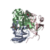
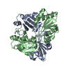
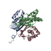
 Novosphingobium aromaticivorans (bacteria)
 X-RAY DIFFRACTION /
  Authors
 Citation
 Structure visualization
 Downloads & links
 Other downloads


 PDBj
PDBj
 Assembly
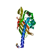
 Mass: 18.015 Da / Num. of mol.: 270 / Source method: isolated from a natural source / Formula: H2O
 Sample preparation
 / Beamline: 8.2.1 / Wavelength: 0.9797, 1.0000
 Processing