 Movie
Movie Controller

[English] 日本語
 Yorodumi
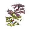
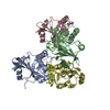
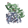
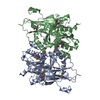
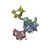
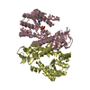
- PDB-2rfk: Substrate RNA Positioning in the Archaeal H/ACA Ribonucleoprotein... -
+ Open data

- Basic information
| Entry | Database: PDB / ID: 2rfk | ||||||
|---|---|---|---|---|---|---|---|
| Title | Substrate RNA Positioning in the Archaeal H/ACA Ribonucleoprotein Complex | ||||||
 Components Components |
| ||||||
 Keywords Keywords | Isomerase/RNA / PROTEIN-RNA COMPLEX / Archaeal H-ACA Ribonucleoprotein Complex / Isomerase / tRNA processing / Ribosome biogenesis / rRNA processing / Isomerase-RNA COMPLEX / Structural Genomics / Southeast Collaboratory for Structural Genomics / SECSG / PSI-2 / Protein Structure Initiative | ||||||
| Function / homology |  Function and homology information Function and homology informationtRNA pseudouridine55 synthase / tRNA pseudouridine(55) synthase activity / pseudouridine synthesis / rRNA pseudouridine synthesis / box H/ACA sno(s)RNA 3'-end processing / tRNA pseudouridine synthesis / snRNA pseudouridine synthesis / mRNA pseudouridine synthesis / snoRNA binding / rRNA processing ...tRNA pseudouridine55 synthase / tRNA pseudouridine(55) synthase activity / pseudouridine synthesis / rRNA pseudouridine synthesis / box H/ACA sno(s)RNA 3'-end processing / tRNA pseudouridine synthesis / snRNA pseudouridine synthesis / mRNA pseudouridine synthesis / snoRNA binding / rRNA processing / ribosome biogenesis / ribonucleoprotein complex / RNA binding Similarity search - Function | ||||||
| Biological species |   Pyrococcus furiosus (archaea) Pyrococcus furiosus (archaea) | ||||||
| Method |  X-RAY DIFFRACTION / SYNCHROTRON / MOLECULAR REPLACEMENT / Resolution: 2.87 Å X-RAY DIFFRACTION / SYNCHROTRON / MOLECULAR REPLACEMENT / Resolution: 2.87 Å | ||||||
 Authors Authors | Liang, B. / Xue, S. / Terns, R.M. / Terns, M.P. / Li, H. / Southeast Collaboratory for Structural Genomics (SECSG) | ||||||
 Citation Citation | Journal: Nat.Struct.Mol.Biol. / Year: 2007 Title: Substrate RNA positioning in the archaeal H/ACA ribonucleoprotein complex. Authors: Liang, B. / Xue, S. / Terns, R.M. / Terns, M.P. / Li, H. | ||||||
| History |
|
- Structure visualization
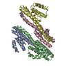
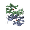
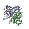
| Structure viewer | Molecule: MolmilJmol/JSmol |
|---|
- Downloads & links
-Download
| PDBx/mmCIF format | 2rfk.cif.gz | 140.7 KB | Display | PDBx/mmCIF format |
|---|---|---|---|---|
| PDB format | pdb2rfk.ent.gz | 104.7 KB | Display | PDB format |
| PDBx/mmJSON format | 2rfk.json.gz | Tree view | PDBx/mmJSON format | |
| Others |  Other downloads Other downloads |
-Validation report
| Arichive directory | https://data.pdbj.org/pub/pdb/validation_reports/rf/2rfkftp://data.pdbj.org/pub/pdb/validation_reports/rf/2rfk | HTTPS FTP |
|---|
-Related structure data
| Related structure data | 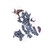 2ey4S S: Starting model for refinement |
|---|---|
| Similar structure data | |
| Other databases |

-Links
 PDBj
PDBj


- Assembly
| Deposited unit | 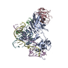
| ||||||||
|---|---|---|---|---|---|---|---|---|---|
| 1 |
| ||||||||
| Unit cell |
|
-Components
-RNA chain , 3 types, 3 molecules DEF
| #1: RNA chain | Mass: 6797.132 Da / Num. of mol.: 1 / Source method: obtained synthetically |
|---|---|
| #2: RNA chain | Mass: 8229.945 Da / Num. of mol.: 1 / Source method: obtained synthetically |
| #3: RNA chain | Mass: 4535.716 Da / Num. of mol.: 1 / Source method: obtained synthetically |
-Protein , 3 types, 3 molecules ABC
| #4: Protein | Mass: 37862.188 Da / Num. of mol.: 1 / Mutation: D85A Source method: isolated from a genetically manipulated source Source: (gene. exp.) Pyrococcus furiosus (archaea) / Plasmid: pET / Production host:  References: UniProt: Q7LWY0, Isomerases; Intramolecular transferases; Transferring other groups |
|---|---|
| #5: Protein | Mass: 6352.509 Da / Num. of mol.: 1 Source method: isolated from a genetically manipulated source Source: (gene. exp.) Pyrococcus furiosus (archaea) / Plasmid: pET / Production host: |
| #6: Protein | Mass: 8515.109 Da / Num. of mol.: 1 Source method: isolated from a genetically manipulated source Source: (gene. exp.) Pyrococcus furiosus (archaea) / Plasmid: pET / Production host: |
-Non-polymers , 1 types, 1 molecules 
| #7: Chemical | ChemComp-ZN /  Mass: 65.409 Da / Num. of mol.: 1 / Source method: obtained synthetically / Formula: Zn Mass: 65.409 Da / Num. of mol.: 1 / Source method: obtained synthetically / Formula: Zn |
|---|
-Details
| Has protein modification | Y |
|---|
-Experimental details
-Experiment
| Experiment | Method: X-RAY DIFFRACTION / Number of used crystals: 1 |
|---|
- Sample preparation
| Crystal | Density Matthews: 3.89 Å3/Da / Density % sol: 68.34 % | ||||||||||||||||||||||||||||||||||||
|---|---|---|---|---|---|---|---|---|---|---|---|---|---|---|---|---|---|---|---|---|---|---|---|---|---|---|---|---|---|---|---|---|---|---|---|---|---|
| Crystal grow | Temperature: 303 K / Method: vapor diffusion, hanging drop / pH: 6 Details: 50 mM MES, 100 mM NH4COOCH3, 5 mM MgSO4, 1.0 M NaCl, pH 6.0, VAPOR DIFFUSION, HANGING DROP, temperature 303K | ||||||||||||||||||||||||||||||||||||
| Components of the solutions |
|
-Data collection
| Diffraction | Mean temperature: 100 K |
|---|---|
| Diffraction source | Source: SYNCHROTRON / Site: APS  / Beamline: 22-ID / Wavelength: 1 Å / Beamline: 22-ID / Wavelength: 1 Å |
| Detector | Type: MARMOSAIC 300 mm CCD / Detector: CCD / Date: Feb 18, 2007 |
| Radiation | Protocol: SINGLE WAVELENGTH / Monochromatic (M) / Laue (L): M / Scattering type: x-ray |
| Radiation wavelength | Wavelength: 1 Å / Relative weight: 1 |
| Reflection | Resolution: 2.87→42.52 Å / Num. all: 29512 / Num. obs: 20534 / % possible obs: 100 % / Observed criterion σ(F): 0 / Observed criterion σ(I): 5 / Redundancy: 15.1 % / Rsym value: 0.11 / Net I/σ(I): 41.9 |
| Reflection shell | Resolution: 2.87→2.945 Å / % possible all: 100 |
- Processing
| Software |
| ||||||||||||||||||||||||||||||||||||||||||||||||||||||||||||||||||||||||||||||||||||||||||||||||||||||||||||||||||||||||||||||||||||||||||||||||||||||||||||||||||||||||||
|---|---|---|---|---|---|---|---|---|---|---|---|---|---|---|---|---|---|---|---|---|---|---|---|---|---|---|---|---|---|---|---|---|---|---|---|---|---|---|---|---|---|---|---|---|---|---|---|---|---|---|---|---|---|---|---|---|---|---|---|---|---|---|---|---|---|---|---|---|---|---|---|---|---|---|---|---|---|---|---|---|---|---|---|---|---|---|---|---|---|---|---|---|---|---|---|---|---|---|---|---|---|---|---|---|---|---|---|---|---|---|---|---|---|---|---|---|---|---|---|---|---|---|---|---|---|---|---|---|---|---|---|---|---|---|---|---|---|---|---|---|---|---|---|---|---|---|---|---|---|---|---|---|---|---|---|---|---|---|---|---|---|---|---|---|---|---|---|---|---|---|---|
| Refinement | Method to determine structure: MOLECULAR REPLACEMENT Starting model: PDB ENTRY 2EY4 Resolution: 2.87→42.52 Å / Cor.coef. Fo:Fc: 0.929 / Cor.coef. Fo:Fc free: 0.91 / SU B: 56.708 / SU ML: 0.46 / Cross valid method: THROUGHOUT / ESU R: 2.43 / ESU R Free: 0.466 / Stereochemistry target values: MAXIMUM LIKELIHOOD / Details: HYDROGENS HAVE BEEN ADDED IN THE RIDING POSITIONS
| ||||||||||||||||||||||||||||||||||||||||||||||||||||||||||||||||||||||||||||||||||||||||||||||||||||||||||||||||||||||||||||||||||||||||||||||||||||||||||||||||||||||||||
| Solvent computation | Ion probe radii: 0.8 Å / Shrinkage radii: 0.8 Å / VDW probe radii: 1.2 Å / Solvent model: BABINET MODEL WITH MASK | ||||||||||||||||||||||||||||||||||||||||||||||||||||||||||||||||||||||||||||||||||||||||||||||||||||||||||||||||||||||||||||||||||||||||||||||||||||||||||||||||||||||||||
| Displacement parameters | Biso mean: 48.255 Å2
| ||||||||||||||||||||||||||||||||||||||||||||||||||||||||||||||||||||||||||||||||||||||||||||||||||||||||||||||||||||||||||||||||||||||||||||||||||||||||||||||||||||||||||
| Refinement step | Cycle: LAST / Resolution: 2.87→42.52 Å
| ||||||||||||||||||||||||||||||||||||||||||||||||||||||||||||||||||||||||||||||||||||||||||||||||||||||||||||||||||||||||||||||||||||||||||||||||||||||||||||||||||||||||||
| Refine LS restraints |
| ||||||||||||||||||||||||||||||||||||||||||||||||||||||||||||||||||||||||||||||||||||||||||||||||||||||||||||||||||||||||||||||||||||||||||||||||||||||||||||||||||||||||||
| LS refinement shell | Resolution: 2.87→2.945 Å / Total num. of bins used: 20
|
