 Movie
Movie Controller


+ Open data

- Basic information
| Entry | Database: PDB / ID: 2rcj | ||||||
|---|---|---|---|---|---|---|---|
| Title | Solution structure of human Immunoglobulin M | ||||||
 Components Components |
| ||||||
 Keywords Keywords | IMMUNE SYSTEM / polymeric antibodies / immunology / constrained modelling | ||||||
| Function / homology |  Function and homology information Function and homology informationcomplement-dependent cytotoxicity / antibody-dependent cellular cytotoxicity / Fc-gamma receptor I complex binding / immunoglobulin complex, circulating / Classical antibody-mediated complement activation / immunoglobulin receptor binding / Initial triggering of complement / IgG immunoglobulin complex / FCGR activation / complement activation, classical pathway ...complement-dependent cytotoxicity / antibody-dependent cellular cytotoxicity / Fc-gamma receptor I complex binding / immunoglobulin complex, circulating / Classical antibody-mediated complement activation / immunoglobulin receptor binding / Initial triggering of complement / IgG immunoglobulin complex / FCGR activation / complement activation, classical pathway / Role of phospholipids in phagocytosis / immunoglobulin complex / antigen binding / FCGR3A-mediated IL10 synthesis / Regulation of Complement cascade / B cell receptor signaling pathway / FCGR3A-mediated phagocytosis / Regulation of actin dynamics for phagocytic cup formation / antibacterial humoral response / Interleukin-4 and Interleukin-13 signaling / blood microparticle / adaptive immune response / : / extracellular exosome / extracellular region / plasma membrane Similarity search - Function | ||||||
| Biological species |  Homo sapiens (human) Homo sapiens (human) | ||||||
| Method |  SOLUTION SCATTERING SOLUTION SCATTERING | ||||||
 Authors Authors | Perkins, S.J. / Nealis, A.S. / Sutton, B.J. / Feinstein, A. | ||||||
 Citation Citation | Journal: J.Mol.Biol. / Year: 1991 Title: Solution structure of human and mouse immunoglobulin M by synchrotron X-ray scattering and molecular graphics modelling. A possible mechanism for complement activation. Authors: Perkins, S.J. / Nealis, A.S. / Sutton, B.J. / Feinstein, A. #1: Journal: J.Immunol. / Year: 2008Title: Implications of the near-planar solution structure of human myeloma dimeric IgA1 for mucosal immunity and IgA nephropathy Authors: Bonner, A. / Furtado, P.B. / Almogren, A. / Kerr, M.A. / Perkins, S.J. | ||||||
| History |
|
- Structure visualization
| Structure viewer | Molecule: MolmilJmol/JSmol |
|---|
- Downloads & links
-Download
| PDBx/mmCIF format | 2rcj.cif.gz | 216.9 KB | Display | PDBx/mmCIF format |
|---|---|---|---|---|
| PDB format | pdb2rcj.ent.gz | 138.7 KB | Display | PDB format |
| PDBx/mmJSON format | 2rcj.json.gz | Tree view | PDBx/mmJSON format | |
| Others |  Other downloads Other downloads |
-Validation report
| Arichive directory | https://data.pdbj.org/pub/pdb/validation_reports/rc/2rcjftp://data.pdbj.org/pub/pdb/validation_reports/rc/2rcj | HTTPS FTP |
|---|
-Related structure data
| Related structure data | |
|---|---|
| Similar structure data |


-Links
 PDBj
PDBj


- Assembly
| Deposited unit | 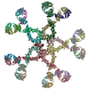
|
|---|---|
| 1 |
|
| Details | This structure is a pentamer structure with 4 chains in each subunit - plus an additional J Chain to make it 21 polypeptide chains. |
-Components
| #1: Antibody | Mass: 23165.770 Da / Num. of mol.: 10 / Source method: isolated from a natural source Details: Human immunoglobulin M was purified from the serum of a patient with Waldenstrom's disease Source: (natural) Homo sapiens (human) / References: UniProt: Q6PYX1*PLUS#2: Antibody | Mass: 56759.504 Da / Num. of mol.: 10 / Source method: isolated from a natural source Details: Human immunoglobulin M was purified from the serum of a patient with Waldenstrom's disease Source: (natural) Homo sapiens (human) / References: UniProt: P01857*PLUS#3: Protein | | Mass: 10700.027 Da / Num. of mol.: 1 / Source method: isolated from a natural source Details: Human immunoglobulin M was purified from the serum of a patient with Waldenstrom's disease Source: (natural) Homo sapiens (human) |
|---|
-Experimental details
-Experiment
| Experiment | Method: SOLUTION SCATTERING |
|---|
-Data collection
| Diffraction |
| ||||||||||||||||||
|---|---|---|---|---|---|---|---|---|---|---|---|---|---|---|---|---|---|---|---|
| Diffraction source |
| ||||||||||||||||||
| Detector |
| ||||||||||||||||||
| Radiation |
| ||||||||||||||||||
| Radiation wavelength | Wavelength: 1.54 Å / Relative weight: 1 | ||||||||||||||||||
| Soln scatter | Type: x-ray / Buffer name: 200 MM NACL 12 MM NAHPO4 0.05% NA AZIDE / Conc. range: 2-30 / Data analysis software list: SCTPL2 / Data reduction software list: SWANAL / Detector type: LINEAR DETECTOR / Max mean cross sectional radii gyration: 1.79 nm / Max mean cross sectional radii gyration esd: 0.12 nm / Mean guiner radius: 12.17 nm / Mean guiner radius esd: 0.34 nm / Min mean cross sectional radii gyration: 6.06 nm / Min mean cross sectional radii gyration esd: 0.19 nm / Num. of time frames: 1 / Sample pH: 7 / Source beamline: 7.3 / Source class: Y / Source type: SRS DARESBURY / Temperature: 293 K |
- Processing
| Software |
| ||||||||||||||
|---|---|---|---|---|---|---|---|---|---|---|---|---|---|---|---|
| Refinement step | Cycle: LAST
| ||||||||||||||
| Soln scatter model | Details: THE DEPOSITED STRUCTURE CONTAINS ONLY CA ATOMS. THE IGM COORDINATES ARE PROVIDED FROM AN EARLY STUDY IN WHICH A MANUAL FIT METHOD BASED ON KNOWN CRYSTAL STRUCTURES FOR IGG WAS USED TO ...Details: THE DEPOSITED STRUCTURE CONTAINS ONLY CA ATOMS. THE IGM COORDINATES ARE PROVIDED FROM AN EARLY STUDY IN WHICH A MANUAL FIT METHOD BASED ON KNOWN CRYSTAL STRUCTURES FOR IGG WAS USED TO DETERMINE STRUCTURES FOR INTACT IGM AND ITS FAB, FAB'2, FC5 AND IGM-S MONOMER SUBUNITS. THESE CRYSTAL STRUCTURES ORIGINATED FROM HUMAN FAB KOL (PDB CODE 2FB4) AND HUMAN FC (PDB CODE 1FC1). THIS PROCEDURE IS APPROXIMATE AND THE IGM MODEL IS NOT CONSIDERED TO BE OPTIMAL OR REFINED AGAINST THE X-RAY SCATTERING DATA. Num. of conformers submitted: 1 / Software list: FRODO, INSIGHT, SCTPL2 |
