 Movie
Movie Controller

[English] 日本語
 Yorodumi
- PDB-2ra8: Crystal structure of the Q64V53_BACFR protein from Bacteroides fr... -
+ Open data

- Basic information
| Entry | Database: PDB / ID: 2ra8 | ||||||
|---|---|---|---|---|---|---|---|
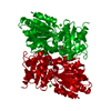
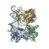
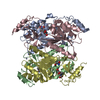
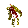
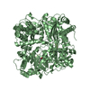
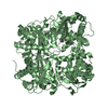
| Title | Crystal structure of the Q64V53_BACFR protein from Bacteroides fragilis. Northeast Structural Genomics Consortium target BfR43 | ||||||
 Components Components | Uncharacterized protein Q64V53_BACFR | ||||||
 Keywords Keywords | STRUCTURAL GENOMICS / UNKNOWN FUNCTION / Q64V53 / WGR DOMAIN / LRR DOMAIN / LEUCINE RICH REPEATS / BFR43 / NESG / PSI-2 / Protein Structure Initiative / Northeast Structural Genomics Consortium | ||||||
| Function / homology |  Function and homology information Function and homology informationWGR domain / : / : / q64v53_bacfr protein fold / WGR domain / WGR domain superfamily / WGR domain / WGR domain profile. / Proposed nucleic acid binding domain / Leucine-rich repeat, LRR (right-handed beta-alpha superhelix) ...WGR domain / : / : / q64v53_bacfr protein fold / WGR domain / WGR domain superfamily / WGR domain / WGR domain profile. / Proposed nucleic acid binding domain / Leucine-rich repeat, LRR (right-handed beta-alpha superhelix) / Ribonuclease Inhibitor / Alpha-Beta Horseshoe / Single Sheet / Leucine-rich repeat domain superfamily / Mainly Beta / Alpha Beta Similarity search - Domain/homology | ||||||
| Biological species |  Bacteroides fragilis (bacteria) Bacteroides fragilis (bacteria) | ||||||
| Method |  X-RAY DIFFRACTION / SYNCHROTRON / SAD / Resolution: 1.95 Å X-RAY DIFFRACTION / SYNCHROTRON / SAD / Resolution: 1.95 Å | ||||||
 Authors Authors | Vorobiev, S.M. / Abashidze, M. / Seetharaman, J. / Wang, D. / Cunningham, K. / Maglaqui, M. / Owens, L. / Xiao, R. / Acton, T.B. / Montelione, G.T. ...Vorobiev, S.M. / Abashidze, M. / Seetharaman, J. / Wang, D. / Cunningham, K. / Maglaqui, M. / Owens, L. / Xiao, R. / Acton, T.B. / Montelione, G.T. / Hunt, J.F. / Tong, L. / Northeast Structural Genomics Consortium (NESG) | ||||||
 Citation Citation | Journal: To be Published Title: Crystal structure of the Q64V53_BACFR protein from Bacteroides fragilis. Authors: Vorobiev, S.M. / Abashidze, M. / Seetharaman, J. / Wang, D. / Cunningham, K. / Maglaqui, M. / Owens, L. / Xiao, R. / Acton, T.B. / Montelione, G.T. / Hunt, J.F. / Tong, L. | ||||||
| History |
|
- Structure visualization
| Structure viewer | Molecule: MolmilJmol/JSmol |
|---|
- Downloads & links
-Download
| PDBx/mmCIF format | 2ra8.cif.gz | 88.2 KB | Display | PDBx/mmCIF format |
|---|---|---|---|---|
| PDB format | pdb2ra8.ent.gz | 65.6 KB | Display | PDB format |
| PDBx/mmJSON format | 2ra8.json.gz | Tree view | PDBx/mmJSON format | |
| Others |  Other downloads Other downloads |
-Validation report
| Arichive directory | https://data.pdbj.org/pub/pdb/validation_reports/ra/2ra8ftp://data.pdbj.org/pub/pdb/validation_reports/ra/2ra8 | HTTPS FTP |
|---|
-Related structure data
| Similar structure data | |
|---|---|
| Other databases |


-Links
 PDBj
PDBj- Assembly
| Deposited unit | 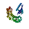
| ||||||||
|---|---|---|---|---|---|---|---|---|---|
| 1 | 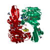
| ||||||||
| 2 |
| ||||||||
| Unit cell |
| ||||||||
| Components on special symmetry positions |
| ||||||||
| Details | Dimer according to the aggregation analysis; second part of the biological assembly is generated by -x+1,y,-z+1 symmetry operator. |
-Components
| #1: Protein | Mass: 41852.574 Da / Num. of mol.: 1 Source method: isolated from a genetically manipulated source Source: (gene. exp.) Bacteroides fragilis (bacteria) / Strain: YCH46 / Gene: BF1877 / Plasmid: pET21-23C / Production host: |
|---|---|
| #2: Water | ChemComp-HOH /  Mass: 18.015 Da / Num. of mol.: 230 / Source method: isolated from a natural source / Formula: H2O Mass: 18.015 Da / Num. of mol.: 230 / Source method: isolated from a natural source / Formula: H2O |
| Has protein modification | Y |
-Experimental details
-Experiment
| Experiment | Method: X-RAY DIFFRACTION / Number of used crystals: 1 |
|---|
- Sample preparation
| Crystal | Density Matthews: 2.36 Å3/Da / Density % sol: 47.91 % Description: The structure factor file contains Friedel pairs |
|---|---|
| Crystal grow | Temperature: 291 K / Method: vapor diffusion, hanging drop / pH: 6.5 Details: 16-18% PEG 3350, 0.2M Magnesium acetate, 0.1M Cacodylic acid pH 6.5, VAPOR DIFFUSION, HANGING DROP, temperature 291K |
-Data collection
| Diffraction | Mean temperature: 100 K |
|---|---|
| Diffraction source | Source: SYNCHROTRON / Site: NSLS  / Beamline: X4A / Wavelength: 0.97922 Å / Beamline: X4A / Wavelength: 0.97922 Å |
| Detector | Type: ADSC QUANTUM 4 / Detector: CCD / Date: Jul 25, 2007 |
| Radiation | Protocol: SINGLE WAVELENGTH / Monochromatic (M) / Laue (L): M / Scattering type: x-ray |
| Radiation wavelength | Wavelength: 0.97922 Å / Relative weight: 1 |
| Reflection | Resolution: 1.95→50 Å / Num. all: 52396 / Num. obs: 52396 / % possible obs: 94.3 % / Observed criterion σ(F): 0 / Observed criterion σ(I): 0 / Redundancy: 1.9 % / Biso Wilson estimate: 9.6 Å2 / Rmerge(I) obs: 0.058 / Net I/σ(I): 14.35 |
| Reflection shell | Resolution: 1.95→2.02 Å / Redundancy: 1.9 % / Rmerge(I) obs: 0.239 / Mean I/σ(I) obs: 2.25 / Num. unique all: 5541 / % possible all: 99.2 |
- Processing
| Software |
| ||||||||||||||||||||||||||||||||||||||||||||||||||||||||||||
|---|---|---|---|---|---|---|---|---|---|---|---|---|---|---|---|---|---|---|---|---|---|---|---|---|---|---|---|---|---|---|---|---|---|---|---|---|---|---|---|---|---|---|---|---|---|---|---|---|---|---|---|---|---|---|---|---|---|---|---|---|---|
| Refinement | Method to determine structure: SAD / Resolution: 1.95→19.39 Å / Rfactor Rfree error: 0.006 / Data cutoff high absF: 622181.64 / Data cutoff low absF: 0 / Isotropic thermal model: RESTRAINED / Cross valid method: THROUGHOUT / σ(F): 2 / Stereochemistry target values: Engh & Huber Details: 1. The Friedel pairs were used for phasing. 2. Cys 116 was modified to s-(dimethylarsenic)cysteine by cacodylic buffer. 3. Tyr 316 is located in a generously allowed position on Ramachandran ...Details: 1. The Friedel pairs were used for phasing. 2. Cys 116 was modified to s-(dimethylarsenic)cysteine by cacodylic buffer. 3. Tyr 316 is located in a generously allowed position on Ramachandran plot. However, Tyr 316 electron density is very well defined and could be interpreted unambiguously.
| ||||||||||||||||||||||||||||||||||||||||||||||||||||||||||||
| Solvent computation | Solvent model: FLAT MODEL / Bsol: 60.8472 Å2 / ksol: 0.348442 e/Å3 | ||||||||||||||||||||||||||||||||||||||||||||||||||||||||||||
| Displacement parameters | Biso mean: 30.1 Å2
| ||||||||||||||||||||||||||||||||||||||||||||||||||||||||||||
| Refine analyze |
| ||||||||||||||||||||||||||||||||||||||||||||||||||||||||||||
| Refinement step | Cycle: LAST / Resolution: 1.95→19.39 Å
| ||||||||||||||||||||||||||||||||||||||||||||||||||||||||||||
| Refine LS restraints |
| ||||||||||||||||||||||||||||||||||||||||||||||||||||||||||||
| LS refinement shell | Resolution: 1.95→2.07 Å / Rfactor Rfree error: 0.021 / Total num. of bins used: 6
| ||||||||||||||||||||||||||||||||||||||||||||||||||||||||||||
| Xplor file |
|
