 Movie
Movie Controller


+ Open data

- Basic information
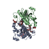
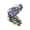
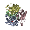
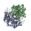
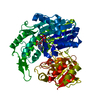
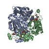
| Entry | Database: PDB / ID: 2r96 | ||||||
|---|---|---|---|---|---|---|---|
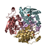
| Title | Crystal structure of E. coli WrbA in complex with FMN | ||||||
 Components Components | Flavoprotein WrbA | ||||||
 Keywords Keywords | OXIDOREDUCTASE / ELECTRON TRANSPORT / quinone oxidoreductase / flavoprotein / flavodoxin-like fold / FMN-binding | ||||||
| Function / homology |  Function and homology information Function and homology informationNAD(P)H dehydrogenase (quinone) / NADPH dehydrogenase (quinone) activity / NADH dehydrogenase (quinone) (non-electrogenic) activity / NAD(P)H dehydrogenase (quinone) activity / NAD binding / NADP binding / FMN binding / flavin adenine dinucleotide binding / response to oxidative stress / protein-containing complex ...NAD(P)H dehydrogenase (quinone) / NADPH dehydrogenase (quinone) activity / NADH dehydrogenase (quinone) (non-electrogenic) activity / NAD(P)H dehydrogenase (quinone) activity / NAD binding / NADP binding / FMN binding / flavin adenine dinucleotide binding / response to oxidative stress / protein-containing complex / membrane / identical protein binding / cytosol Similarity search - Function | ||||||
| Biological species |  | ||||||
| Method |  X-RAY DIFFRACTION / SYNCHROTRON / MOLECULAR REPLACEMENT / molecular replacement / Resolution: 2.6 Å X-RAY DIFFRACTION / SYNCHROTRON / MOLECULAR REPLACEMENT / molecular replacement / Resolution: 2.6 Å | ||||||
 Authors Authors | Kuta Smatanova, I. / Wolfova, J. / Brynda, J. / Mesters, J.R. / Grandori, R. / Carey, J. | ||||||
 Citation Citation | Journal: Biochim.Biophys.Acta / Year: 2009 Title: Structural organization of WrbA in apo- and holoprotein crystals. Authors: Wolfova, J. / Smatanova, I.K. / Brynda, J. / Mesters, J.R. / Lapkouski, M. / Kuty, M. / Natalello, A. / Chatterjee, N. / Chern, S.Y. / Ebbel, E. / Ricci, A. / Grandori, R. / Ettrich, R. / Carey, J. #1: Journal: Protein Sci. / Year: 2007Title: WrbA bridges bacterial flavodoxins and eukaryotic NAD(P)H:quinone oxidoreductases Authors: Carey, J. / Brynda, J. / Wolfova, J. / Grandori, R. / Gustavsson, T. / Ettrich, R. / Kuta Smatanova, I. #2: Journal: Acta Crystallogr.,Sect.F / Year: 2007Title: Crystallization and preliminary diffraction analysis of Escherichia coli WrbA in complex with its cofactor flavin mononucleotide Authors: Wolfova, J. / Mesters, J.R. / Brynda, J. / Grandori, R. / Natalello, A. / Carey, J. / Kuta Smatanova, I. | ||||||
| History |
|
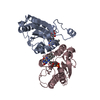
- Structure visualization
| Structure viewer | Molecule: MolmilJmol/JSmol |
|---|
- Downloads & links
-Download
| PDBx/mmCIF format | 2r96.cif.gz | 91.2 KB | Display | PDBx/mmCIF format |
|---|---|---|---|---|
| PDB format | pdb2r96.ent.gz | 69.4 KB | Display | PDB format |
| PDBx/mmJSON format | 2r96.json.gz | Tree view | PDBx/mmJSON format | |
| Others |  Other downloads Other downloads |
-Validation report
| Arichive directory | https://data.pdbj.org/pub/pdb/validation_reports/r9/2r96ftp://data.pdbj.org/pub/pdb/validation_reports/r9/2r96 | HTTPS FTP |
|---|
-Related structure data
| Related structure data |  2r97C  2rg1C  1zwlS C: citing same article ( S: Starting model for refinement |
|---|---|
| Similar structure data |

-Links
 PDBj
PDBj

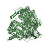
- Assembly
| Deposited unit | 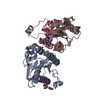
| ||||||||||||||||||||||||||||||||||||||||||||||||||||||||||
|---|---|---|---|---|---|---|---|---|---|---|---|---|---|---|---|---|---|---|---|---|---|---|---|---|---|---|---|---|---|---|---|---|---|---|---|---|---|---|---|---|---|---|---|---|---|---|---|---|---|---|---|---|---|---|---|---|---|---|---|
| 1 | 
| ||||||||||||||||||||||||||||||||||||||||||||||||||||||||||
| Unit cell |
| ||||||||||||||||||||||||||||||||||||||||||||||||||||||||||
| Noncrystallographic symmetry (NCS) | NCS domain:
NCS domain segments: Component-ID: 1 / Beg auth comp-ID: ALA / Beg label comp-ID: ALA / Refine code: 4
NCS ensembles :
| ||||||||||||||||||||||||||||||||||||||||||||||||||||||||||
| Details | The biological assembly is a tetramer generated from the dimer in the asymmetric unit by the operations: -y, -x, 1/2-z. |
-Components
| #1: Protein | Mass: 20862.473 Da / Num. of mol.: 2 Source method: isolated from a genetically manipulated source Source: (gene. exp.) References: UniProt: P0A8G6, NAD(P)H dehydrogenase (quinone) #2: Chemical |   Mass: 456.344 Da / Num. of mol.: 2 / Source method: obtained synthetically / Formula: C17H21N4O9P Mass: 456.344 Da / Num. of mol.: 2 / Source method: obtained synthetically / Formula: C17H21N4O9P#3: Chemical | ChemComp-EDO /   Mass: 62.068 Da / Num. of mol.: 14 / Source method: obtained synthetically / Formula: C2H6O2 Mass: 62.068 Da / Num. of mol.: 14 / Source method: obtained synthetically / Formula: C2H6O2#4: Water | ChemComp-HOH / |  Mass: 18.015 Da / Num. of mol.: 101 / Source method: isolated from a natural source / Formula: H2O Mass: 18.015 Da / Num. of mol.: 101 / Source method: isolated from a natural source / Formula: H2O |
|---|
-Experimental details
-Experiment
| Experiment | Method: X-RAY DIFFRACTION / Number of used crystals: 1 |
|---|
- Sample preparation
| Crystal | Density Matthews: 4.68 Å3/Da / Density % sol: 73.7 % |
|---|---|
| Crystal grow | Temperature: 285 K / Method: vapor diffusion, sitting drop / pH: 7.5 Details: 25% ethylene glycol, VAPOR DIFFUSION, SITTING DROP, temperature 285K, pH 7.5 |
-Data collection
| Diffraction | Mean temperature: 100 K |
|---|---|
| Diffraction source | Source: SYNCHROTRON / Site: EMBL/DESY, HAMBURG  / Beamline: X13 / Wavelength: 0.81 Å / Beamline: X13 / Wavelength: 0.81 Å |
| Detector | Type: MAR CCD 165 mm / Detector: CCD / Date: May 10, 2006 / Details: mirrors |
| Radiation | Monochromator: Si (111), horizontally focussing / Protocol: SINGLE WAVELENGTH / Monochromatic (M) / Laue (L): M / Scattering type: x-ray |
| Radiation wavelength | Wavelength: 0.81 Å / Relative weight: 1 |
| Reflection | Resolution: 2.6→35 Å / Num. all: 25285 / Num. obs: 24986 / % possible obs: 98.9 % / Redundancy: 7.2 % / Limit h max: 36 / Limit h min: 0 / Limit k max: 25 / Limit k min: 0 / Limit l max: 67 / Limit l min: 0 / Rmerge(I) obs: 0.14 / Net I/σ(I): 12.35 |
| Reflection shell | Resolution: 2.6→2.69 Å / Redundancy: 6.7 % / Rmerge(I) obs: 0.62 / Mean I/σ(I) obs: 2.64 / Num. unique all: 2435 / % possible all: 98.1 |
-Phasing
| Phasing | Method: molecular replacement |
|---|
- Processing
| Software |
| ||||||||||||||||||||||||||||||||||||||||||||||||||||||||||||||||||||||||||||||||||||||||||
|---|---|---|---|---|---|---|---|---|---|---|---|---|---|---|---|---|---|---|---|---|---|---|---|---|---|---|---|---|---|---|---|---|---|---|---|---|---|---|---|---|---|---|---|---|---|---|---|---|---|---|---|---|---|---|---|---|---|---|---|---|---|---|---|---|---|---|---|---|---|---|---|---|---|---|---|---|---|---|---|---|---|---|---|---|---|---|---|---|---|---|---|
| Refinement | Method to determine structure: MOLECULAR REPLACEMENT Starting model: PDB ENTRY 1ZWL Resolution: 2.6→34.28 Å / Cor.coef. Fo:Fc: 0.933 / Cor.coef. Fo:Fc free: 0.904 / Isotropic thermal model: Isotropic / Cross valid method: THROUGHOUT / σ(F): 0 / ESU R: 0.28 / ESU R Free: 0.228 / Stereochemistry target values: MAXIMUM LIKELIHOOD
| ||||||||||||||||||||||||||||||||||||||||||||||||||||||||||||||||||||||||||||||||||||||||||
| Solvent computation | Ion probe radii: 0.8 Å / Shrinkage radii: 0.8 Å / VDW probe radii: 1.4 Å / Solvent model: MASK | ||||||||||||||||||||||||||||||||||||||||||||||||||||||||||||||||||||||||||||||||||||||||||
| Displacement parameters | Biso mean: 29.112 Å2
| ||||||||||||||||||||||||||||||||||||||||||||||||||||||||||||||||||||||||||||||||||||||||||
| Refinement step | Cycle: LAST / Resolution: 2.6→34.28 Å
| ||||||||||||||||||||||||||||||||||||||||||||||||||||||||||||||||||||||||||||||||||||||||||
| Refine LS restraints |
| ||||||||||||||||||||||||||||||||||||||||||||||||||||||||||||||||||||||||||||||||||||||||||
| Refine LS restraints NCS | Refine-ID: X-RAY DIFFRACTION
| ||||||||||||||||||||||||||||||||||||||||||||||||||||||||||||||||||||||||||||||||||||||||||
| LS refinement shell | Resolution: 2.6→2.67 Å / Total num. of bins used: 20
|
