 Movie
Movie Controller


+ Open data

- Basic information
| Entry | Database: PDB / ID: 2r91 | ||||||
|---|---|---|---|---|---|---|---|
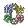
| Title | Crystal Structure of KD(P)GA from T.tenax | ||||||
 Components Components | 2-Keto-3-deoxy-(6-phospho-)gluconate aldolase | ||||||
 Keywords Keywords | LYASE / TIM barrel / aldolase / thermophilic | ||||||
| Function / homology |  Function and homology information Function and homology information2-dehydro-3-deoxy-D-gluconate aldolase / 2-dehydro-3-deoxy-D-gluconate aldolase activity / 2-dehydro-3-deoxy-phosphogluconate aldolase / 2-dehydro-3-deoxy-phosphogluconate aldolase activity / 4-hydroxy-tetrahydrodipicolinate synthase activity Similarity search - Function | ||||||
| Biological species |   Thermoproteus tenax (archaea) Thermoproteus tenax (archaea) | ||||||
| Method |  X-RAY DIFFRACTION / SYNCHROTRON / Resolution: 2 Å X-RAY DIFFRACTION / SYNCHROTRON / Resolution: 2 Å | ||||||
 Authors Authors | Pauluhn, A. / Pohl, E. / Lorentzen, E. / Siebers, B. / Ahmed, H. / Buchinger, S. / Schomburg, D. | ||||||
 Citation Citation | Journal: Proteins / Year: 2008 Title: Crystal structure and stereochemical studies of KD(P)G aldolase from Thermoproteus tenax. Authors: Pauluhn, A. / Ahmed, H. / Lorentzen, E. / Buchinger, S. / Schomburg, D. / Siebers, B. / Pohl, E. | ||||||
| History |
|
- Structure visualization
| Structure viewer | Molecule: MolmilJmol/JSmol |
|---|
- Downloads & links
-Download
| PDBx/mmCIF format | 2r91.cif.gz | 235.1 KB | Display | PDBx/mmCIF format |
|---|---|---|---|---|
| PDB format | pdb2r91.ent.gz | 190.3 KB | Display | PDB format |
| PDBx/mmJSON format | 2r91.json.gz | Tree view | PDBx/mmJSON format | |
| Others |  Other downloads Other downloads |
-Validation report
| Summary document | 2r91_validation.pdf.gz | 453.1 KB | Display | wwPDB validaton report |
|---|---|---|---|---|
| Full document | 2r91_full_validation.pdf.gz | 467.4 KB | Display | |
| Data in XML | 2r91_validation.xml.gz | 48.9 KB | Display | |
| Data in CIF | 2r91_validation.cif.gz | 70.3 KB | Display | |
| Arichive directory | https://data.pdbj.org/pub/pdb/validation_reports/r9/2r91ftp://data.pdbj.org/pub/pdb/validation_reports/r9/2r91 | HTTPS FTP |
-Related structure data

-Links
 PDBj
PDBj- Assembly
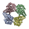
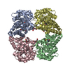
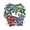
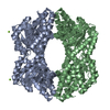
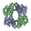
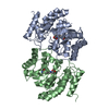
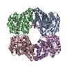
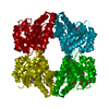
| Deposited unit | 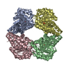
| ||||||||||||||||||||||||||||||
|---|---|---|---|---|---|---|---|---|---|---|---|---|---|---|---|---|---|---|---|---|---|---|---|---|---|---|---|---|---|---|---|
| 1 |
| ||||||||||||||||||||||||||||||
| Unit cell |
| ||||||||||||||||||||||||||||||
| Noncrystallographic symmetry (NCS) | NCS domain:
NCS domain segments: Component-ID: 1 / Ens-ID: 1 / Beg auth comp-ID: MET / Beg label comp-ID: MET / End auth comp-ID: LEU / End label comp-ID: LEU / Refine code: 4 / Auth seq-ID: 21 - 306 / Label seq-ID: 1 - 286
|
-Components
| #1: Protein | Mass: 31017.344 Da / Num. of mol.: 4 Source method: isolated from a genetically manipulated source Source: (gene. exp.) Thermoproteus tenax (archaea) / Gene: kdgA / Plasmid: pET-15b / Species (production host): Escherichia coli / Production host:  References: UniProt: Q704D1, Lyases; Carbon-carbon lyases; Aldehyde-lyases #2: Chemical | ChemComp-SO4 /   Mass: 96.063 Da / Num. of mol.: 4 / Source method: obtained synthetically / Formula: SO4 Mass: 96.063 Da / Num. of mol.: 4 / Source method: obtained synthetically / Formula: SO4#3: Water | ChemComp-HOH / |  Mass: 18.015 Da / Num. of mol.: 690 / Source method: isolated from a natural source / Formula: H2O Mass: 18.015 Da / Num. of mol.: 690 / Source method: isolated from a natural source / Formula: H2OHas protein modification | Y | |
|---|
-Experimental details
-Experiment
| Experiment | Method: X-RAY DIFFRACTION / Number of used crystals: 1 |
|---|
- Sample preparation
| Crystal | Density Matthews: 3.99 Å3/Da / Density % sol: 69.21 % |
|---|---|
| Crystal grow | Temperature: 298 K / Method: vapor diffusion / pH: 7 Details: HEPES/KOH, pH 7.0, VAPOR DIFFUSION, temperature 298K |
-Data collection
| Diffraction | Mean temperature: 100 K |
|---|---|
| Diffraction source | Source: SYNCHROTRON / Site: SLS  / Beamline: X10SA / Wavelength: 0.905 Å / Beamline: X10SA / Wavelength: 0.905 Å |
| Detector | Type: MARMOSAIC 225 mm CCD / Detector: CCD / Date: Apr 20, 2005 |
| Radiation | Monochromator: Si(111) / Protocol: SINGLE WAVELENGTH / Monochromatic (M) / Laue (L): M / Scattering type: x-ray |
| Radiation wavelength | Wavelength: 0.905 Å / Relative weight: 1 |
| Reflection | Resolution: 2→48.17 Å / Num. all: 135342 / Num. obs: 135046 / Observed criterion σ(I): 3 / Redundancy: 9.9 % / Rmerge(I) obs: 0.065 / Rsym value: 0.087 |
| Reflection shell | Resolution: 2→2.1 Å / Redundancy: 10 % / Rmerge(I) obs: 0.25 / Mean I/σ(I) obs: 5.92 / Num. unique all: 18158 / Rsym value: 0.392 / % possible all: 100 |
- Processing
| Software |
| ||||||||||||||||||||||||||||||||||||||||||||||||||||||||||||||||||||||||||||||||||||||||||
|---|---|---|---|---|---|---|---|---|---|---|---|---|---|---|---|---|---|---|---|---|---|---|---|---|---|---|---|---|---|---|---|---|---|---|---|---|---|---|---|---|---|---|---|---|---|---|---|---|---|---|---|---|---|---|---|---|---|---|---|---|---|---|---|---|---|---|---|---|---|---|---|---|---|---|---|---|---|---|---|---|---|---|---|---|---|---|---|---|---|---|---|
| Refinement | Resolution: 2→48.17 Å / Cor.coef. Fo:Fc: 0.954 / Cor.coef. Fo:Fc free: 0.939 / SU B: 2.644 / SU ML: 0.076 / Cross valid method: THROUGHOUT / σ(F): 0 / ESU R: 0.121 / ESU R Free: 0.118 / Stereochemistry target values: MAXIMUM LIKELIHOOD / Details: HYDROGENS HAVE BEEN ADDED IN THE RIDING POSITIONS
| ||||||||||||||||||||||||||||||||||||||||||||||||||||||||||||||||||||||||||||||||||||||||||
| Solvent computation | Ion probe radii: 0.8 Å / Shrinkage radii: 0.8 Å / VDW probe radii: 1.4 Å / Solvent model: MASK | ||||||||||||||||||||||||||||||||||||||||||||||||||||||||||||||||||||||||||||||||||||||||||
| Displacement parameters | Biso mean: 27.08 Å2
| ||||||||||||||||||||||||||||||||||||||||||||||||||||||||||||||||||||||||||||||||||||||||||
| Refinement step | Cycle: LAST / Resolution: 2→48.17 Å
| ||||||||||||||||||||||||||||||||||||||||||||||||||||||||||||||||||||||||||||||||||||||||||
| Refine LS restraints |
| ||||||||||||||||||||||||||||||||||||||||||||||||||||||||||||||||||||||||||||||||||||||||||
| Refine LS restraints NCS | Ens-ID: 1 / Number: 2185 / Refine-ID: X-RAY DIFFRACTION
| ||||||||||||||||||||||||||||||||||||||||||||||||||||||||||||||||||||||||||||||||||||||||||
| LS refinement shell | Resolution: 2→2.052 Å / Total num. of bins used: 20
|
