 Movie
Movie Controller

[English] 日本語
 Yorodumi
- PDB-2r5r: The crystal structure of DUF198 from Nitrosomonas europaea ATCC 19718 -
+ Open data

- Basic information
| Entry | Database: PDB / ID: 2r5r | ||||||
|---|---|---|---|---|---|---|---|
| Title | The crystal structure of DUF198 from Nitrosomonas europaea ATCC 19718 | ||||||
 Components Components | UPF0343 protein NE1163 | ||||||
 Keywords Keywords | STRUCTURAL GENOMICS / UNKNOWN FUNCTION / APC86493 / DUF198 / Nitrosomonas europaea ATCC 19718 / PSI-2 / Protein Structure Initiative / Midwest Center for Structural Genomics / MCSG | ||||||
| Function / homology |  Function and homology information Function and homology informationGTP cyclohydrolase I / GTP cyclohydrolase I activity / tetrahydrofolate biosynthetic process Similarity search - Function | ||||||
| Biological species |  Nitrosomonas europaea ATCC 19718 (bacteria) Nitrosomonas europaea ATCC 19718 (bacteria) | ||||||
| Method |  X-RAY DIFFRACTION / SYNCHROTRON / MAD / Resolution: 3.05 Å X-RAY DIFFRACTION / SYNCHROTRON / MAD / Resolution: 3.05 Å | ||||||
 Authors Authors | Tan, K. / Wu, R. / Nocek, B. / Bigelow, L. / Patterson, S. / Freeman, L. / Bargassa, M. / Joachimiak, A. / Midwest Center for Structural Genomics (MCSG) | ||||||
 Citation Citation | Journal: To be Published Title: The crystal structure of DUF198 from Nitrosomonas europaea ATCC 19718. Authors: Tan, K. / Wu, R. / Nocek, B. / Bigelow, L. / Patterson, S. / Freeman, L. / Bargassa, M. / Joachimiak, A. | ||||||
| History |
|
- Structure visualization
| Structure viewer | Molecule: MolmilJmol/JSmol |
|---|
- Downloads & links
-Download
| PDBx/mmCIF format | 2r5r.cif.gz | 62.3 KB | Display | PDBx/mmCIF format |
|---|---|---|---|---|
| PDB format | pdb2r5r.ent.gz | 45.5 KB | Display | PDB format |
| PDBx/mmJSON format | 2r5r.json.gz | Tree view | PDBx/mmJSON format | |
| Others |  Other downloads Other downloads |
-Validation report
| Arichive directory | https://data.pdbj.org/pub/pdb/validation_reports/r5/2r5rftp://data.pdbj.org/pub/pdb/validation_reports/r5/2r5r | HTTPS FTP |
|---|
-Related structure data
| Similar structure data | |
|---|---|
| Other databases |


-Links
 PDBj
PDBj


- Assembly
| Deposited unit | 
| ||||||||
|---|---|---|---|---|---|---|---|---|---|
| 1 | 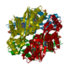
| ||||||||
| Unit cell |
| ||||||||
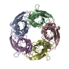
| Details | Authors state that the biological unit is experimentally unknown. From molecular packing, it seems to be a tetramer, generated from symmetry operators given in remark 350. |
-Components
| #1: Protein | Mass: 31201.350 Da / Num. of mol.: 1 Source method: isolated from a genetically manipulated source Source: (gene. exp.) Nitrosomonas europaea ATCC 19718 (bacteria)Species: Nitrosomonas europaea / Strain: IFO 14298 / Gene: NE1163 / Plasmid: pMCSG7 / Species (production host): Escherichia coli / Production host: |
|---|---|
| #2: Chemical | ChemComp-PO4 /   Mass: 94.971 Da / Num. of mol.: 1 / Source method: obtained synthetically / Formula: PO4 Mass: 94.971 Da / Num. of mol.: 1 / Source method: obtained synthetically / Formula: PO4 |
| #3: Chemical | ChemComp-IMD /   Mass: 69.085 Da / Num. of mol.: 1 / Source method: obtained synthetically / Formula: C3H5N2 Mass: 69.085 Da / Num. of mol.: 1 / Source method: obtained synthetically / Formula: C3H5N2 |
| Has protein modification | Y |
-Experimental details
-Experiment
| Experiment | Method: X-RAY DIFFRACTION / Number of used crystals: 1 |
|---|
- Sample preparation
| Crystal | Density Matthews: 3.69 Å3/Da / Density % sol: 66.66 % |
|---|---|
| Crystal grow | Temperature: 289 K / Method: vapor diffusion, sitting drop / pH: 4.2 Details: 1.6M NaH2PO4/0.4M K2HPO4, 0.1M Phosphate-Citrate, pH 4.2, VAPOR DIFFUSION, SITTING DROP, temperature 289K |
-Data collection
| Diffraction | Mean temperature: 100 K | |||||||||
|---|---|---|---|---|---|---|---|---|---|---|
| Diffraction source | Source: SYNCHROTRON / Site: APS  / Beamline: 19-ID / Wavelength: 0.97951, 0.97965 / Beamline: 19-ID / Wavelength: 0.97951, 0.97965 | |||||||||
| Detector | Type: ADSC QUANTUM 315 / Detector: CCD / Date: Jul 30, 2007 / Details: mirror | |||||||||
| Radiation | Monochromator: Si 111 crystal / Protocol: MAD / Monochromatic (M) / Laue (L): M / Scattering type: x-ray | |||||||||
| Radiation wavelength |
| |||||||||
| Reflection | Resolution: 3.05→38.87 Å / Num. all: 9363 / Num. obs: 9363 / % possible obs: 99.1 % / Observed criterion σ(F): 0 / Observed criterion σ(I): 0 / Redundancy: 9.9 % / Rmerge(I) obs: 0.151 / Net I/σ(I): 17.4 | |||||||||
| Reflection shell | Resolution: 3.05→3.16 Å / Redundancy: 7.9 % / Rmerge(I) obs: 0.847 / Mean I/σ(I) obs: 1.6 / Num. unique all: 846 / % possible all: 93.6 |
- Processing
| Software |
| ||||||||||||||||||||||||||||||||||||||||||||||||||||||||||||||||||||||||||||||||||||||||||||||||||||||||||||||||||||||||||||||||||||||||||||||||||||||||||||||||||||||||||
|---|---|---|---|---|---|---|---|---|---|---|---|---|---|---|---|---|---|---|---|---|---|---|---|---|---|---|---|---|---|---|---|---|---|---|---|---|---|---|---|---|---|---|---|---|---|---|---|---|---|---|---|---|---|---|---|---|---|---|---|---|---|---|---|---|---|---|---|---|---|---|---|---|---|---|---|---|---|---|---|---|---|---|---|---|---|---|---|---|---|---|---|---|---|---|---|---|---|---|---|---|---|---|---|---|---|---|---|---|---|---|---|---|---|---|---|---|---|---|---|---|---|---|---|---|---|---|---|---|---|---|---|---|---|---|---|---|---|---|---|---|---|---|---|---|---|---|---|---|---|---|---|---|---|---|---|---|---|---|---|---|---|---|---|---|---|---|---|---|---|---|---|
| Refinement | Method to determine structure: MAD / Resolution: 3.05→38.87 Å / Cor.coef. Fo:Fc: 0.915 / Cor.coef. Fo:Fc free: 0.902 / SU B: 17.744 / SU ML: 0.307 / Cross valid method: THROUGHOUT / σ(F): 0 / σ(I): 0 / ESU R: 1.083 / ESU R Free: 0.383 / Stereochemistry target values: MAXIMUM LIKELIHOOD / Details: HYDROGENS HAVE BEEN ADDED IN THE RIDING POSITIONS
| ||||||||||||||||||||||||||||||||||||||||||||||||||||||||||||||||||||||||||||||||||||||||||||||||||||||||||||||||||||||||||||||||||||||||||||||||||||||||||||||||||||||||||
| Solvent computation | Ion probe radii: 0.8 Å / Shrinkage radii: 0.8 Å / VDW probe radii: 1.2 Å / Solvent model: MASK | ||||||||||||||||||||||||||||||||||||||||||||||||||||||||||||||||||||||||||||||||||||||||||||||||||||||||||||||||||||||||||||||||||||||||||||||||||||||||||||||||||||||||||
| Displacement parameters | Biso mean: 74.918 Å2
| ||||||||||||||||||||||||||||||||||||||||||||||||||||||||||||||||||||||||||||||||||||||||||||||||||||||||||||||||||||||||||||||||||||||||||||||||||||||||||||||||||||||||||
| Refinement step | Cycle: LAST / Resolution: 3.05→38.87 Å
| ||||||||||||||||||||||||||||||||||||||||||||||||||||||||||||||||||||||||||||||||||||||||||||||||||||||||||||||||||||||||||||||||||||||||||||||||||||||||||||||||||||||||||
| Refine LS restraints |
| ||||||||||||||||||||||||||||||||||||||||||||||||||||||||||||||||||||||||||||||||||||||||||||||||||||||||||||||||||||||||||||||||||||||||||||||||||||||||||||||||||||||||||
| LS refinement shell | Resolution: 3.05→3.13 Å / Total num. of bins used: 20
|
