 Movie
Movie Controller

[English] 日本語
 Yorodumi
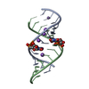
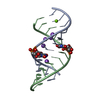
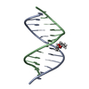
- PDB-2r22: Structure of the native RNA tridecamer r(GCGUUUGAAACGC) at 1.5 A ... -
+ Open data

- Basic information
| Entry | Database: PDB / ID: 2r22 | ||||||||||||||||||
|---|---|---|---|---|---|---|---|---|---|---|---|---|---|---|---|---|---|---|---|
| Title | Structure of the native RNA tridecamer r(GCGUUUGAAACGC) at 1.5 A (NatMn) | ||||||||||||||||||
 Components Components | RNA (5'-R(* Keywords KeywordsRNA / monovalent / electrostatic / asymmetry / manganese | Function / homology | : / RNA / RNA (> 10) |  Function and homology information Function and homology informationMethod |  X-RAY DIFFRACTION / SYNCHROTRON / Resolution: 1.4 Å X-RAY DIFFRACTION / SYNCHROTRON / Resolution: 1.4 Å  Authors AuthorsTimsit, Y. / Bombard, S. |  CitationJournal: TO BE PUBLISHED CitationJournal: TO BE PUBLISHEDTitle: The 1.3 A structure of the tridecamer r(GCGUUUGAAACGC) Authors: Timsit, Y. / Bombard, S. History |
|
- Structure visualization
| Structure viewer | Molecule: MolmilJmol/JSmol |
|---|
- Downloads & links
-Download
| PDBx/mmCIF format | 2r22.cif.gz | 27.9 KB | Display | PDBx/mmCIF format |
|---|---|---|---|---|
| PDB format | pdb2r22.ent.gz | 18.2 KB | Display | PDB format |
| PDBx/mmJSON format | 2r22.json.gz | Tree view | PDBx/mmJSON format | |
| Others |  Other downloads Other downloads |
-Validation report
| Arichive directory | https://data.pdbj.org/pub/pdb/validation_reports/r2/2r22ftp://data.pdbj.org/pub/pdb/validation_reports/r2/2r22 | HTTPS FTP |
|---|
-Related structure data
| Related structure data | |
|---|---|
| Similar structure data |


-Links
 PDBj
PDBj


- Assembly
| Deposited unit | 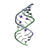
| ||||||||
|---|---|---|---|---|---|---|---|---|---|
| 1 |
| ||||||||
| Unit cell |
|
-Components
| #1: RNA chain | Mass: 4157.525 Da / Num. of mol.: 2 / Source method: obtained synthetically #2: Chemical | ChemComp-NA /   Mass: 22.990 Da / Num. of mol.: 4 / Source method: obtained synthetically / Formula: Na Mass: 22.990 Da / Num. of mol.: 4 / Source method: obtained synthetically / Formula: Na#3: Chemical | ChemComp-MN / |   Mass: 54.938 Da / Num. of mol.: 1 / Source method: obtained synthetically / Formula: Mn Mass: 54.938 Da / Num. of mol.: 1 / Source method: obtained synthetically / Formula: Mn#4: Water | ChemComp-HOH / |  Mass: 18.015 Da / Num. of mol.: 145 / Source method: isolated from a natural source / Formula: H2O Mass: 18.015 Da / Num. of mol.: 145 / Source method: isolated from a natural source / Formula: H2O |
|---|
-Experimental details
-Experiment
| Experiment | Method: X-RAY DIFFRACTION / Number of used crystals: 1 |
|---|
- Sample preparation
| Crystal | Density Matthews: 2.11 Å3/Da / Density % sol: 41.58 % | ||||||||||||||||||||||||||||||||||||
|---|---|---|---|---|---|---|---|---|---|---|---|---|---|---|---|---|---|---|---|---|---|---|---|---|---|---|---|---|---|---|---|---|---|---|---|---|---|
| Crystal grow | Method: vapor diffusion, hanging drop / pH: 7 Details: 3 mM MnCl2, 100 mM NaCl, 50 mM Tris, 30 % MPD, pH 7.0, VAPOR DIFFUSION, HANGING DROP | ||||||||||||||||||||||||||||||||||||
| Components of the solutions |
|
-Data collection
| Diffraction | Mean temperature: 80 K |
|---|---|
| Diffraction source | Source: SYNCHROTRON / Site: ESRF  / Beamline: BM30A / Wavelength: 0.919487 Å / Beamline: BM30A / Wavelength: 0.919487 Å |
| Detector | Type: MAR scanner 345 mm plate / Detector: IMAGE PLATE / Date: Feb 3, 2003 / Details: mirrors |
| Radiation | Monochromator: Si (111) Crystal / Protocol: SINGLE WAVELENGTH / Monochromatic (M) / Laue (L): M / Scattering type: x-ray |
| Radiation wavelength | Wavelength: 0.919487 Å / Relative weight: 1 |
| Reflection | Resolution: 1.4→20 Å / Num. all: 12814 / Num. obs: 12814 / % possible obs: 88.7 % / Redundancy: 1.7 % / Rmerge(I) obs: 0.09 / Rsym value: 6.4 / Net I/σ(I): 7.8 |
| Reflection shell | Resolution: 1.4→1.53 Å / Redundancy: 1.1 % / Rmerge(I) obs: 0.279 / Mean I/σ(I) obs: 2.1 / Num. unique all: 1838 / Rsym value: 19.7 / % possible all: 92 |
- Processing
| Software |
| ||||||||||||||||||||||||||||
|---|---|---|---|---|---|---|---|---|---|---|---|---|---|---|---|---|---|---|---|---|---|---|---|---|---|---|---|---|---|
| Refinement | Resolution: 1.4→20 Å / σ(F): 104
| ||||||||||||||||||||||||||||
| Solvent computation | Bsol: 40.159 Å2 | ||||||||||||||||||||||||||||
| Displacement parameters | Biso mean: 13.727 Å2
| ||||||||||||||||||||||||||||
| Refinement step | Cycle: LAST / Resolution: 1.4→20 Å
| ||||||||||||||||||||||||||||
| Refine LS restraints |
| ||||||||||||||||||||||||||||
| Xplor file |
|
