Entry Database : PDB / ID : 2r1tTitle dopamine quinone conjugation to DJ-1 (DJ-1) x 2 Keywords / / / / / / / / / / / / / / Function / homology Function Domain/homology Component
/ / / / / / / / / / / / / / / / / / / / / / / / / / / / / / / / / / / / / / / / / / / / / / / / / / / / / / / / / / / / / / / / / / / / / / / / / / / / / / / / / / / / / / / / / / / / / / / / / / / / / / / / / / / / / / / / / / / / Biological species Homo sapiens (human)Method / / / / Resolution : 1.7 Å Authors Zhongtao, Z. / Yue, F. Journal : To be Published Title : DJ-1 is a proisopeptidaseAuthors : Zhongtao, Z. / Yue, F. History Deposition Aug 23, 2007 Deposition site / Processing site Revision 1.0 Aug 26, 2008 Provider / Type Revision 1.1 Jul 13, 2011 Group Revision 1.2 Oct 25, 2017 Group / Source and taxonomy / Category / softwareItem / _entity_src_gen.pdbx_host_org_ncbi_taxonomy_id / _software.nameRevision 1.3 Oct 20, 2021 Group / Derived calculationsCategory database_2 / struct_conn ... database_2 / struct_conn / struct_ref_seq_dif / struct_sheet Item _database_2.pdbx_DOI / _database_2.pdbx_database_accession ... _database_2.pdbx_DOI / _database_2.pdbx_database_accession / _struct_conn.pdbx_leaving_atom_flag / _struct_ref_seq_dif.details / _struct_sheet.number_strands
Show all Show less
 Movie
Movie Controller


 Open data
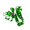
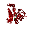
 Basic information
 Components
 Keywords
 Function and homology information
 Homo sapiens (human)
 X-RAY DIFFRACTION /
  Authors
 Citation
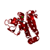
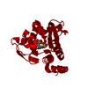
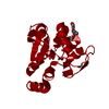
 Structure visualization
 Downloads & links
 Other downloads


 PDBj
PDBj


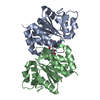
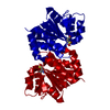
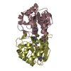
 Assembly
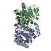

 Mass: 18.015 Da / Num. of mol.: 417 / Source method: isolated from a natural source / Formula: H2O
 Sample preparation
 / Beamline: 19-BM / Wavelength: 1 Å
 Processing