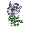
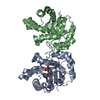
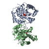
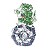
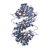
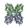
BIOMOLECULE: 1, 2 SEE REMARK 350 FOR THE AUTHOR PROVIDED AND PROGRAM GENERATED ASSEMBLY ... BIOMOLECULE: 1, 2 SEE REMARK 350 FOR THE AUTHOR PROVIDED AND PROGRAM GENERATED ASSEMBLY INFORMATION FOR THE STRUCTURE IN THIS ENTRY. THE REMARK MAY ALSO PROVIDE INFORMATION ON BURIED SURFACE AREA. SIZE EXCLUSION CHROMATOGRAPHY SUPPORTS THE ASSIGNMENT OF A DIMER AS THE SIGNIFICANT OLIGOMERIZATION STATE.
Remark 999
SEQUENCE THE CONSTRUCT WAS EXPRESSED WITH A PURIFICATION TAG MGSDKIHHHHHHENLYFQG. THE TAG WAS ... SEQUENCE THE CONSTRUCT WAS EXPRESSED WITH A PURIFICATION TAG MGSDKIHHHHHHENLYFQG. THE TAG WAS REMOVED WITH TEV PROTEASE LEAVING ONLY A GLYCINE (0) FOLLOWED BY THE TARGET SEQUENCE.
Resolution: 1.5→38.152 Å / Num. obs: 87321 / % possible obs: 99.8 % / Redundancy: 7.7 % / Rmerge(I) obs: 0.057 / Rsym value: 0.057 / Net I/σ(I): 8.7
Reflection shell
Diffraction-ID: 1
Resolution (Å)
Redundancy (%)
Rmerge(I) obs
Mean I/σ(I) obs
Num. measured all
Num. unique all
Rsym value
% possible all
1.5-1.54
4.8
0.688
0.9
29805
6228
0.688
98.1
1.54-1.58
6
0.63
1.2
37336
6219
0.63
99.9
1.58-1.63
7.2
0.553
1.2
43669
6046
0.553
100
1.63-1.68
7.2
0.465
1.6
42567
5892
0.465
100
1.68-1.73
7.2
0.37
2.1
41229
5691
0.37
100
1.73-1.79
7.2
0.273
2.8
39985
5526
0.273
100
1.79-1.86
7.2
0.221
3.5
38737
5353
0.221
100
1.86-1.94
7.2
0.161
4.1
37230
5140
0.161
100
1.94-2.02
7.2
0.135
5.3
35912
4963
0.135
100
2.02-2.12
7.2
0.124
4.4
33875
4715
0.124
100
2.12-2.24
7.2
0.106
5.8
32482
4517
0.106
100
2.24-2.37
7.2
0.093
5.8
30885
4282
0.093
100
2.37-2.54
7.9
0.081
7.4
32041
4057
0.081
100
2.54-2.74
10.8
0.069
9.2
40219
3735
0.069
100
2.74-3
10.7
0.057
10.5
37652
3507
0.057
100
3-3.35
10.7
0.044
13.4
33590
3150
0.044
100
3.35-3.87
10.5
0.036
16.5
30066
2853
0.036
100
3.87-4.74
10.3
0.029
20.6
24922
2410
0.029
100
4.74-6.71
10
0.027
20.8
19148
1916
0.027
100
6.71-38.152
8.5
0.025
23.2
9552
1121
0.025
98.3
-
Phasing
Phasing
Method: MAD
-
Processing
Software
Name
Version
Classification
NB
REFMAC
5.2.0019
refinement
PHENIX
refinement
SHELX
phasing
MolProbity
3beta29
modelbuilding
SCALA
datascaling
PDB_EXTRACT
2
dataextraction
ADSC
Quantum
datacollection
MOSFLM
datareduction
SHARP
phasing
SHELXD
phasing
Refinement
Method to determine structure: MAD / Resolution: 1.5→38.152 Å / Cor.coef. Fo:Fc: 0.972 / Cor.coef. Fo:Fc free: 0.967 / SU B: 2.342 / SU ML: 0.042 / TLS residual ADP flag: LIKELY RESIDUAL / Cross valid method: THROUGHOUT / σ(F): 0 / ESU R: 0.06 / ESU R Free: 0.06 Stereochemistry target values: MAXIMUM LIKELIHOOD WITH PHASES Details: 1. HYDROGENS HAVE BEEN ADDED IN THE RIDING POSITIONS. 2. ATOM RECORDS CONTAIN RESIDUAL B FACTORS ONLY. 3. A MET-INHIBITION PROTOCOL WAS USED FOR SELENOMETHIONINE INCORPORATION DURING PROTEIN ...Details: 1. HYDROGENS HAVE BEEN ADDED IN THE RIDING POSITIONS. 2. ATOM RECORDS CONTAIN RESIDUAL B FACTORS ONLY. 3. A MET-INHIBITION PROTOCOL WAS USED FOR SELENOMETHIONINE INCORPORATION DURING PROTEIN EXPRESSION. THE OCCUPANCY OF THE SE ATOMS IN THE MSE RESIDUES WAS REDUCED TO 0.75 FOR THE REDUCED SCATTERING POWER DUE TO PARTIAL S-MET INCORPORATION. 4. RESIDUES 1-2, 81-82 IN CHAIN A AND 1-4, 81-85 IN CHAIN B ARE DISORDERED AND NOT INCLUDED IN THE MODEL. 5. IMIDAZOLE MOLECULES FROM THE PURIFICATION SOLUTION ARE MODELED. 6. GLYCEROL MOLECULES FROM THE CRYO SOLUTION ARE MODELED. 7. IMIDAZOLE AND ONE GLYCEROL MOLECULES OCCUPY THE ACTIVE SITE OF THE PROTEIN.
Rfactor
Num. reflection
% reflection
Selection details
Rfree
0.184
4372
5 %
RANDOM
Rwork
0.165
-
-
-
all
0.166
-
-
-
obs
0.166
87230
99.78 %
-
Solvent computation
Ion probe radii: 0.8 Å / Shrinkage radii: 0.8 Å / VDW probe radii: 1.2 Å / Solvent model: BABINET MODEL WITH MASK
In the structure databanks used in Yorodumi, some data are registered as the other names, "COVID-19 virus" and "2019-nCoV". Here are the details of the virus and the list of structure data.
Jan 31, 2019. EMDB accession codes are about to change! (news from PDBe EMDB page)
EMDB accession codes are about to change! (news from PDBe EMDB page)
The allocation of 4 digits for EMDB accession codes will soon come to an end. Whilst these codes will remain in use, new EMDB accession codes will include an additional digit and will expand incrementally as the available range of codes is exhausted. The current 4-digit format prefixed with “EMD-” (i.e. EMD-XXXX) will advance to a 5-digit format (i.e. EMD-XXXXX), and so on. It is currently estimated that the 4-digit codes will be depleted around Spring 2019, at which point the 5-digit format will come into force.
The EM Navigator/Yorodumi systems omit the EMD- prefix.
Related info.:Q: What is EMD? / ID/Accession-code notation in Yorodumi/EM Navigator
Yorodumi is a browser for structure data from EMDB, PDB, SASBDB, etc.
This page is also the successor to EM Navigator detail page, and also detail information page/front-end page for Omokage search.
The word "yorodu" (or yorozu) is an old Japanese word meaning "ten thousand". "mi" (miru) is to see.
Related info.:EMDB / PDB / SASBDB / Comparison of 3 databanks / Yorodumi Search / Aug 31, 2016. New EM Navigator & Yorodumi / Yorodumi Papers / Jmol/JSmol / Function and homology information / Changes in new EM Navigator and Yorodumi
 Movie
Movie Controller

 Yorodumi
 Open data
 Basic information
 Components
 Keywords
 Function and homology information
 Sulfolobus solfataricus P2 (archaea)
 X-RAY DIFFRACTION /
  Authors
 Citation
 Structure visualization
 Downloads & links
 Other downloads
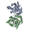


 PDBj
PDBj


 Assembly


 Mass: 69.085 Da / Num. of mol.: 2 / Source method: obtained synthetically / Formula: C3H5N2
 Mass: 92.094 Da / Num. of mol.: 7 / Source method: obtained synthetically / Formula: C3H8O3
 Mass: 18.015 Da / Num. of mol.: 312 / Source method: isolated from a natural source / Formula: H2O
 Sample preparation
 / Beamline: BL1-5 / Wavelength: 0.918381, 0.979310, 0.978921
 Processing