 Movie
Movie Controller

[English] 日本語
 Yorodumi
- PDB-2qyc: Crystal structure of a dimeric ferredoxin-like protein (bb1511) f... -
+ Open data

- Basic information
| Entry | Database: PDB / ID: 2qyc | ||||||
|---|---|---|---|---|---|---|---|
| Title | Crystal structure of a dimeric ferredoxin-like protein (bb1511) from bordetella bronchiseptica rb50 at 1.90 A resolution | ||||||
 Components Components | Ferredoxin-like protein | ||||||
 Keywords Keywords | UNKNOWN FUNCTION / Stress responsive a/b barrel domain / structural genomics / Joint Center for Structural Genomics / JCSG / Protein Structure Initiative / PSI-2 | ||||||
| Function / homology |  Function and homology information Function and homology informationStress responsive alpha-beta barrel / Stress responsive A/B Barrel Domain / Stress-response A/B barrel domain profile. / Stress responsive A/B Barrel Domain / Alpha-Beta Plaits - #100 / Dimeric alpha-beta barrel / Alpha-Beta Plaits / 2-Layer Sandwich / Alpha Beta Similarity search - Domain/homology | ||||||
| Biological species |  Bordetella bronchiseptica RB50 (bacteria) Bordetella bronchiseptica RB50 (bacteria) | ||||||
| Method |  X-RAY DIFFRACTION / SYNCHROTRON / MAD / Resolution: 1.9 Å X-RAY DIFFRACTION / SYNCHROTRON / MAD / Resolution: 1.9 Å | ||||||
 Authors Authors | Joint Center for Structural Genomics (JCSG) | ||||||
 Citation Citation | Journal: To be published Title: Crystal structure of dimeric ferredoxin-like protein (NP_888056.1) from Bordetella bronchiseptica at 1.90 A resolution Authors: Joint Center for Structural Genomics (JCSG) | ||||||
| History |
| ||||||
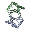
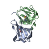
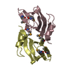
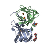
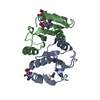
| Remark 300 | BIOMOLECULE: 1, 2 SEE REMARK 350 FOR THE AUTHOR PROVIDED AND PROGRAM GENERATED ASSEMBLY ... BIOMOLECULE: 1, 2 SEE REMARK 350 FOR THE AUTHOR PROVIDED AND PROGRAM GENERATED ASSEMBLY INFORMATION FOR THE STRUCTURE IN THIS ENTRY. THE REMARK MAY ALSO PROVIDE INFORMATION ON BURIED SURFACE AREA. CRYSTAL STRUCTURE PACKING SUGGESTS THAT THE BIOLOGICALLY RELEVANT FORM IS A DIMER. SIZE EXCLUSION CHROMATOGRAPHY SUPPORTS THE ASSIGNMENT OF A DIMER AS A SIGNIFICANT OLIGOMERIZATION STATE IN SOLUTION. | ||||||
| Remark 999 | SEQUENCE THE CONSTRUCT WAS EXPRESSED WITH A PURIFICATION TAG MGSDKIHHHHHHENLYFQG. THE TAG WAS ... SEQUENCE THE CONSTRUCT WAS EXPRESSED WITH A PURIFICATION TAG MGSDKIHHHHHHENLYFQG. THE TAG WAS REMOVED WITH TEV PROTEASE LEAVING ONLY A GLYCINE (0) FOLLOWED BY THE TARGET SEQUENCE. |
- Structure visualization
| Structure viewer | Molecule: MolmilJmol/JSmol |
|---|
- Downloads & links
-Download
| PDBx/mmCIF format | 2qyc.cif.gz | 58.1 KB | Display | PDBx/mmCIF format |
|---|---|---|---|---|
| PDB format | pdb2qyc.ent.gz | 42.4 KB | Display | PDB format |
| PDBx/mmJSON format | 2qyc.json.gz | Tree view | PDBx/mmJSON format | |
| Others |  Other downloads Other downloads |
-Validation report
| Arichive directory | https://data.pdbj.org/pub/pdb/validation_reports/qy/2qycftp://data.pdbj.org/pub/pdb/validation_reports/qy/2qyc | HTTPS FTP |
|---|
-Related structure data
| Similar structure data | |
|---|---|
| Other databases |


-Links
 PDBj
PDBj- Assembly
| Deposited unit | 
| |||||||||
|---|---|---|---|---|---|---|---|---|---|---|
| 1 |
| |||||||||
| 2 | 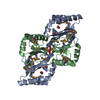
| |||||||||
| Unit cell |
| |||||||||
| Components on special symmetry positions |
| |||||||||
| Details | CRYSTAL STRUCTURE PACKING SUGGESTS THAT THE BIOLOGICALLY RELEVANT FORM IS A DIMER. SIZE EXCLUSION CHROMATOGRAPHY SUPPORTS THE ASSIGNMENT OF A DIMER AS A SIGNIFICANT OLIGOMERIZATION STATE IN SOLUTION. |
-Components
| #1: Protein | Mass: 11722.313 Da / Num. of mol.: 2 Source method: isolated from a genetically manipulated source Source: (gene. exp.) Bordetella bronchiseptica RB50 (bacteria)Species: Bordetella bronchiseptica / Strain: RB50, NCTC 13252 / Gene: NP_888056.1, BB1511 / Plasmid: speedET / Production host: #2: Chemical |   Mass: 62.068 Da / Num. of mol.: 2 / Source method: obtained synthetically / Formula: C2H6O2 Mass: 62.068 Da / Num. of mol.: 2 / Source method: obtained synthetically / Formula: C2H6O2#3: Chemical | ChemComp-CL / |   Mass: 35.453 Da / Num. of mol.: 1 / Source method: obtained synthetically / Formula: Cl Mass: 35.453 Da / Num. of mol.: 1 / Source method: obtained synthetically / Formula: Cl#4: Chemical | ChemComp-ACT / |   Mass: 59.044 Da / Num. of mol.: 1 / Source method: obtained synthetically / Formula: C2H3O2 Mass: 59.044 Da / Num. of mol.: 1 / Source method: obtained synthetically / Formula: C2H3O2#5: Water | ChemComp-HOH / |  Mass: 18.015 Da / Num. of mol.: 120 / Source method: isolated from a natural source / Formula: H2O Mass: 18.015 Da / Num. of mol.: 120 / Source method: isolated from a natural source / Formula: H2OHas protein modification | Y | |
|---|
-Experimental details
-Experiment
| Experiment | Method: X-RAY DIFFRACTION / Number of used crystals: 1 |
|---|
- Sample preparation
| Crystal | Density Matthews: 2.34 Å3/Da / Density % sol: 47.48 % |
|---|---|
| Crystal grow | Temperature: 277 K / Method: vapor diffusion, sitting drop / pH: 4.5 Details: NANODROP, 0.2M Li2SO4, 2.5M NaCl, 0.1M Acetate pH 4.5, VAPOR DIFFUSION, SITTING DROP, temperature 277K |
-Data collection
| Diffraction | Mean temperature: 100 K | ||||||||||||||||||||||||||||||||||||||||||||||||||||||||||||||||||
|---|---|---|---|---|---|---|---|---|---|---|---|---|---|---|---|---|---|---|---|---|---|---|---|---|---|---|---|---|---|---|---|---|---|---|---|---|---|---|---|---|---|---|---|---|---|---|---|---|---|---|---|---|---|---|---|---|---|---|---|---|---|---|---|---|---|---|---|
| Diffraction source | Source: SYNCHROTRON / Site: SSRL  / Beamline: BL11-1 / Wavelength: 0.91837, 0.97926 / Beamline: BL11-1 / Wavelength: 0.91837, 0.97926 | ||||||||||||||||||||||||||||||||||||||||||||||||||||||||||||||||||
| Detector | Type: MARMOSAIC 325 mm CCD / Detector: CCD / Date: Jul 26, 2007 / Details: Flat mirror (vertical focusing) | ||||||||||||||||||||||||||||||||||||||||||||||||||||||||||||||||||
| Radiation | Monochromator: Single crystal Si(111) bent (horizontal focusing) Protocol: MAD / Monochromatic (M) / Laue (L): M / Scattering type: x-ray | ||||||||||||||||||||||||||||||||||||||||||||||||||||||||||||||||||
| Radiation wavelength |
| ||||||||||||||||||||||||||||||||||||||||||||||||||||||||||||||||||
| Reflection | Resolution: 1.9→28.433 Å / Num. obs: 17516 / % possible obs: 99.7 % / Redundancy: 7.04 % / Biso Wilson estimate: 23.22 Å2 / Rmerge(I) obs: 0.087 / Net I/σ(I): 9.74 | ||||||||||||||||||||||||||||||||||||||||||||||||||||||||||||||||||
| Reflection shell |
|
-Phasing
| Phasing | Method: MAD |
|---|
- Processing
| Software |
| |||||||||||||||||||||||||||||||||||||||||||||||||||||||||||||||||||||||||||||||||||||||||||||||||||||||||||||||||||||||||||||
|---|---|---|---|---|---|---|---|---|---|---|---|---|---|---|---|---|---|---|---|---|---|---|---|---|---|---|---|---|---|---|---|---|---|---|---|---|---|---|---|---|---|---|---|---|---|---|---|---|---|---|---|---|---|---|---|---|---|---|---|---|---|---|---|---|---|---|---|---|---|---|---|---|---|---|---|---|---|---|---|---|---|---|---|---|---|---|---|---|---|---|---|---|---|---|---|---|---|---|---|---|---|---|---|---|---|---|---|---|---|---|---|---|---|---|---|---|---|---|---|---|---|---|---|---|---|---|
| Refinement | Method to determine structure: MAD / Resolution: 1.9→28.433 Å / Cor.coef. Fo:Fc: 0.96 / Cor.coef. Fo:Fc free: 0.934 / SU B: 6.788 / SU ML: 0.101 / TLS residual ADP flag: LIKELY RESIDUAL / Cross valid method: THROUGHOUT / σ(F): 0 / ESU R: 0.145 / ESU R Free: 0.148 Stereochemistry target values: MAXIMUM LIKELIHOOD WITH PHASES Details: 1. HYDROGENS HAVE BEEN ADDED IN THE RIDING POSITIONS. 2. A MET-INHIBITION PROTOCOL WAS USED FOR SELENOMETHIONINE INCORPORATION DURING PROTEIN EXPRESSION. THE OCCUPANCY OF THE SE ATOMS IN THE ...Details: 1. HYDROGENS HAVE BEEN ADDED IN THE RIDING POSITIONS. 2. A MET-INHIBITION PROTOCOL WAS USED FOR SELENOMETHIONINE INCORPORATION DURING PROTEIN EXPRESSION. THE OCCUPANCY OF THE SE ATOMS IN THE MSE RESIDUES WAS REDUCED TO 0.75 FOR THE REDUCED SCATTERING POWER DUE TO PARTIAL S-MET INCORPORATION. 3. ATOM RECORD CONTAINS RESIDUAL B FACTORS ONLY. 4. CL, ACETATE, AND ETHYLENE GLYCOL ARE MODELED BASED ON CRYSTALLIZATION AND CRYO CONDITIONS.
| |||||||||||||||||||||||||||||||||||||||||||||||||||||||||||||||||||||||||||||||||||||||||||||||||||||||||||||||||||||||||||||
| Solvent computation | Ion probe radii: 0.8 Å / Shrinkage radii: 0.8 Å / VDW probe radii: 1.2 Å / Solvent model: MASK | |||||||||||||||||||||||||||||||||||||||||||||||||||||||||||||||||||||||||||||||||||||||||||||||||||||||||||||||||||||||||||||
| Displacement parameters | Biso mean: 21.44 Å2
| |||||||||||||||||||||||||||||||||||||||||||||||||||||||||||||||||||||||||||||||||||||||||||||||||||||||||||||||||||||||||||||
| Refinement step | Cycle: LAST / Resolution: 1.9→28.433 Å
| |||||||||||||||||||||||||||||||||||||||||||||||||||||||||||||||||||||||||||||||||||||||||||||||||||||||||||||||||||||||||||||
| Refine LS restraints |
| |||||||||||||||||||||||||||||||||||||||||||||||||||||||||||||||||||||||||||||||||||||||||||||||||||||||||||||||||||||||||||||
| LS refinement shell | Resolution: 1.9→1.949 Å / Total num. of bins used: 20
| |||||||||||||||||||||||||||||||||||||||||||||||||||||||||||||||||||||||||||||||||||||||||||||||||||||||||||||||||||||||||||||
| Refinement TLS params. | Method: refined / Refine-ID: X-RAY DIFFRACTION
| |||||||||||||||||||||||||||||||||||||||||||||||||||||||||||||||||||||||||||||||||||||||||||||||||||||||||||||||||||||||||||||
| Refinement TLS group | Refine-ID: X-RAY DIFFRACTION / Selection: ALL / Auth seq-ID: 0 - 101 / Label seq-ID: 1 - 102
|
