 Movie
Movie Controller


+ Open data

- Basic information
| Entry | Database: PDB / ID: 2qwy | ||||||
|---|---|---|---|---|---|---|---|
| Title | SAM-II riboswitch bound to S-adenosylmethionine | ||||||
 Components Components | SAM-II riboswitch | ||||||
 Keywords Keywords | RNA / mRNA / riboswitch / SAM / S-adenosylmethionine / AdoMet / RNA-ligand complex / double helix / pseudoknot / base triple | ||||||
| Function / homology | : / PHOSPHATE ION / S-ADENOSYLMETHIONINE / RNA / RNA (> 10) Function and homology information Function and homology information | ||||||
| Method |  X-RAY DIFFRACTION / SIRAS / Resolution: 2.8 Å X-RAY DIFFRACTION / SIRAS / Resolution: 2.8 Å | ||||||
 Authors Authors | Gilbert, S.D. / Rambo, R.P. / Van Tyne, D. / Batey, R.T. | ||||||
 Citation Citation | Journal: Nat.Struct.Mol.Biol. / Year: 2008 Title: Structure of the SAM-II riboswitch bound to S-adenosylmethionine. Authors: Gilbert, S.D. / Rambo, R.P. / Van Tyne, D. / Batey, R.T. | ||||||
| History |
|
- Structure visualization
| Structure viewer | Molecule: MolmilJmol/JSmol |
|---|
- Downloads & links
-Download
| PDBx/mmCIF format | 2qwy.cif.gz | 101.1 KB | Display | PDBx/mmCIF format |
|---|---|---|---|---|
| PDB format | pdb2qwy.ent.gz | 77 KB | Display | PDB format |
| PDBx/mmJSON format | 2qwy.json.gz | Tree view | PDBx/mmJSON format | |
| Others |  Other downloads Other downloads |
-Validation report
| Arichive directory | https://data.pdbj.org/pub/pdb/validation_reports/qw/2qwyftp://data.pdbj.org/pub/pdb/validation_reports/qw/2qwy | HTTPS FTP |
|---|
-Related structure data
| Similar structure data |
|---|


-Links
 PDBj
PDBj


- Assembly
| Deposited unit | 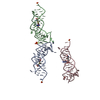
| ||||||||
|---|---|---|---|---|---|---|---|---|---|
| 1 | 
| ||||||||
| 2 | 
| ||||||||
| 3 | 
| ||||||||
| Unit cell |
| ||||||||
| Details | The deposited coordinates contain all information corresponding to one biologically relevant unit repeated three times according to the number of RNA-ligand complexes observed in the asymmetric unit. |
-Components
-RNA chain , 1 types, 3 molecules ABC
| #1: RNA chain | Mass: 16821.043 Da / Num. of mol.: 3 / Source method: obtained synthetically Details: The sequence was engineered based on the SAM-II riboswitch found upstream of the metX gene derived from a Sargasso Sea environmental sequence (Env12). |
|---|
-Non-polymers , 5 types, 166 molecules ABC


| #2: Chemical | ChemComp-PO4 /  Mass: 94.971 Da / Num. of mol.: 6 / Source method: obtained synthetically / Formula: PO4 Mass: 94.971 Da / Num. of mol.: 6 / Source method: obtained synthetically / Formula: PO4#3: Chemical |  Mass: 132.905 Da / Num. of mol.: 2 / Source method: obtained synthetically / Formula: Cs Mass: 132.905 Da / Num. of mol.: 2 / Source method: obtained synthetically / Formula: Cs#4: Chemical | ChemComp-MG / |  Mass: 24.305 Da / Num. of mol.: 1 / Source method: obtained synthetically / Formula: Mg Mass: 24.305 Da / Num. of mol.: 1 / Source method: obtained synthetically / Formula: Mg#5: Chemical |  Mass: 398.437 Da / Num. of mol.: 3 / Source method: obtained synthetically / Formula: C15H22N6O5S Mass: 398.437 Da / Num. of mol.: 3 / Source method: obtained synthetically / Formula: C15H22N6O5S#6: Water | ChemComp-HOH / | Mass: 18.015 Da / Num. of mol.: 154 / Source method: isolated from a natural source / Formula: H2O |
|---|
-Details
| Nonpolymer details | EACH RNA MONOMER CONTAINS A 5'-DIPHOSPHATE AND A 2',3'-CYCLIC PHOSPHATE. THESE RESIDUES HAVE BEEN ...EACH RNA MONOMER CONTAINS A 5'-DIPHOSPHAT |
|---|
-Experimental details
-Experiment
| Experiment | Method: X-RAY DIFFRACTION / Number of used crystals: 1 |
|---|
- Sample preparation
| Crystal | Density Matthews: 2.86 Å3/Da / Density % sol: 56.98 % | ||||||||||||||||||||||||||||||||||||||||||||
|---|---|---|---|---|---|---|---|---|---|---|---|---|---|---|---|---|---|---|---|---|---|---|---|---|---|---|---|---|---|---|---|---|---|---|---|---|---|---|---|---|---|---|---|---|---|
| Crystal grow | Temperature: 298 K / Method: vapor diffusion, hanging drop / pH: 6 Details: 640 mM Ammonium Acetate, 10% PEG 1000, 10 mM Barium Chloride, 50 mM Sodium Cacodylate, 8 mM Cobalt Hexaammine Chloride. Micro-seeded., pH 6.0, VAPOR DIFFUSION, HANGING DROP, temperature 298K | ||||||||||||||||||||||||||||||||||||||||||||
| Components of the solutions |
|
-Data collection
| Diffraction | Mean temperature: 100 K |
|---|---|
| Diffraction source | Source: ROTATING ANODE / Type: RIGAKU / Wavelength: 1.5418 Å |
| Detector | Date: Apr 10, 2007 |
| Radiation | Monochromator: Nickel Filter / Protocol: SINGLE WAVELENGTH / Monochromatic (M) / Laue (L): M / Scattering type: x-ray |
| Radiation wavelength | Wavelength: 1.5418 Å / Relative weight: 1 |
| Reflection | Resolution: 2.8→20 Å / Num. all: 27442 / Num. obs: 26866 / % possible obs: 97.9 % / Observed criterion σ(F): 0 / Observed criterion σ(I): 3 / Redundancy: 7.8 % / Biso Wilson estimate: 129.5 Å2 / Rsym value: 0.061 / Net I/σ(I): 31.9 |
| Reflection shell | Resolution: 2.8→2.9 Å / Redundancy: 7.7 % / Mean I/σ(I) obs: 5 / Num. unique all: 4033 / Rsym value: 0.349 / % possible all: 94.7 |
- Processing
| Software |
| ||||||||||||||||||||||||||||||||||||||||||||||||||||||||||||
|---|---|---|---|---|---|---|---|---|---|---|---|---|---|---|---|---|---|---|---|---|---|---|---|---|---|---|---|---|---|---|---|---|---|---|---|---|---|---|---|---|---|---|---|---|---|---|---|---|---|---|---|---|---|---|---|---|---|---|---|---|---|
| Refinement | Method to determine structure: SIRAS / Resolution: 2.8→19.9 Å / Rfactor Rfree error: 0.006 / Data cutoff high absF: 1022121.53 / Data cutoff low absF: 0 / Isotropic thermal model: RESTRAINED / Cross valid method: THROUGHOUT / σ(F): 0 / σ(I): 2 / Details: BULK SOLVENT MODEL USED
| ||||||||||||||||||||||||||||||||||||||||||||||||||||||||||||
| Solvent computation | Solvent model: FLAT MODEL / Bsol: 9.21319 Å2 / ksol: 0.25 e/Å3 | ||||||||||||||||||||||||||||||||||||||||||||||||||||||||||||
| Displacement parameters | Biso mean: 65.6 Å2
| ||||||||||||||||||||||||||||||||||||||||||||||||||||||||||||
| Refine analyze |
| ||||||||||||||||||||||||||||||||||||||||||||||||||||||||||||
| Refinement step | Cycle: LAST / Resolution: 2.8→19.9 Å
| ||||||||||||||||||||||||||||||||||||||||||||||||||||||||||||
| Refine LS restraints |
| ||||||||||||||||||||||||||||||||||||||||||||||||||||||||||||
| LS refinement shell | Resolution: 2.8→2.97 Å / Rfactor Rfree error: 0.023 / Total num. of bins used: 6
| ||||||||||||||||||||||||||||||||||||||||||||||||||||||||||||
| Xplor file |
|
