 Movie
Movie Controller

[English] 日本語
 Yorodumi
- PDB-2qw6: Crystal structure of the C-terminal domain of an AAA ATPase from ... -
+ Open data

- Basic information
| Entry | Database: PDB / ID: 2qw6 | ||||||
|---|---|---|---|---|---|---|---|
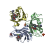
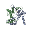
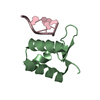
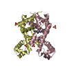
| Title | Crystal structure of the C-terminal domain of an AAA ATPase from Enterococcus faecium DO | ||||||
 Components Components | AAA ATPase, central region | ||||||
 Keywords Keywords | HYDROLASE / STRUCTURAL GENOMICS / ATPASE / PSI-2 / Protein Structure Initiative / New York SGX Research Center for Structural Genomics / NYSGXRC / ATP-binding / Nucleotide-binding | ||||||
| Function / homology | Zinc Finger, Delta Prime; domain 3 - #10 / Zinc Finger, Delta Prime; domain 3 / Up-down Bundle / Mainly Alpha / :  Function and homology information Function and homology information | ||||||
| Biological species |  Enterococcus faecium (bacteria) Enterococcus faecium (bacteria) | ||||||
| Method |  X-RAY DIFFRACTION / SYNCHROTRON / Resolution: 2.3 Å X-RAY DIFFRACTION / SYNCHROTRON / Resolution: 2.3 Å | ||||||
 Authors Authors | Bonanno, J.B. / Rutter, M. / Bain, K.T. / Ozyurt, S. / Smith, D. / Wasserman, S. / Sauder, J.M. / Burley, S.K. / Almo, S.C. / New York SGX Research Center for Structural Genomics (NYSGXRC) | ||||||
 Citation Citation | Journal: To be Published Title: Crystal structure of the C-terminal domain of an AAA ATPase from Enterococcus faecium DO. Authors: Bonanno, J.B. / Rutter, M. / Bain, K.T. / Ozyurt, S. / Smith, D. / Wasserman, S. / Sauder, J.M. / Burley, S.K. / Almo, S.C. | ||||||
| History |
| ||||||
| Remark 300 | BIOMOLECULE: 1 SEE REMARK 350 FOR THE AUTHOR PROVIDED AND PROGRAM GENERATED ASSEMBLY INFORMATION ... BIOMOLECULE: 1 SEE REMARK 350 FOR THE AUTHOR PROVIDED AND PROGRAM GENERATED ASSEMBLY INFORMATION FOR THE STRUCTURE IN THIS ENTRY. THE REMARK MAY ALSO PROVIDE INFORMATION ON BURIED SURFACE AREA. AUTHORS STATE THAT THE TETRAMERIC ASSEMBLY SHOWN IN REMARK 350 IS PROBABLE. |
- Structure visualization
| Structure viewer | Molecule: MolmilJmol/JSmol |
|---|
- Downloads & links
-Download
| PDBx/mmCIF format | 2qw6.cif.gz | 74.6 KB | Display | PDBx/mmCIF format |
|---|---|---|---|---|
| PDB format | pdb2qw6.ent.gz | 56.2 KB | Display | PDB format |
| PDBx/mmJSON format | 2qw6.json.gz | Tree view | PDBx/mmJSON format | |
| Others |  Other downloads Other downloads |
-Validation report
| Arichive directory | https://data.pdbj.org/pub/pdb/validation_reports/qw/2qw6ftp://data.pdbj.org/pub/pdb/validation_reports/qw/2qw6 | HTTPS FTP |
|---|
-Related structure data
| Similar structure data | |
|---|---|
| Other databases |


-Links
 PDBj
PDBj- Assembly
| Deposited unit | 
| ||||||||
|---|---|---|---|---|---|---|---|---|---|
| 1 |
| ||||||||
| Unit cell |
| ||||||||
| Details | probable tetramer |
-Components
| #1: Protein | Mass: 11998.666 Da / Num. of mol.: 4 / Fragment: C-terminal domain: Residues 230-330 Source method: isolated from a genetically manipulated source Source: (gene. exp.) Enterococcus faecium (bacteria) / Strain: DO / Gene: EfaeDRAFT_0938 / Plasmid: Modified pET26 / Species (production host): Escherichia coli / Production host: #2: Water | ChemComp-HOH / |  Mass: 18.015 Da / Num. of mol.: 65 / Source method: isolated from a natural source / Formula: H2O Mass: 18.015 Da / Num. of mol.: 65 / Source method: isolated from a natural source / Formula: H2O |
|---|
-Experimental details
-Experiment
| Experiment | Method: X-RAY DIFFRACTION / Number of used crystals: 1 |
|---|
- Sample preparation
| Crystal | Density Matthews: 2.5 Å3/Da / Density % sol: 50.78 % |
|---|---|
| Crystal grow | Temperature: 294 K / Method: vapor diffusion / pH: 6.5 Details: 100mM Imidazole pH 6.5, 1M Sodium acetate hydrate, VAPOR DIFFUSION, temperature 294K |
-Data collection
| Diffraction | Mean temperature: 100 K |
|---|---|
| Diffraction source | Source: SYNCHROTRON / Site: APS  / Beamline: 31-ID / Wavelength: 0.97958 Å / Beamline: 31-ID / Wavelength: 0.97958 Å |
| Detector | Type: MAR CCD 165 mm / Detector: CCD / Date: Jul 29, 2007 |
| Radiation | Monochromator: diamond / Protocol: SINGLE WAVELENGTH / Monochromatic (M) / Laue (L): M / Scattering type: x-ray |
| Radiation wavelength | Wavelength: 0.97958 Å / Relative weight: 1 |
| Reflection | Resolution: 2.3→87.706 Å / Num. all: 22041 / Num. obs: 22041 / % possible obs: 100 % / Observed criterion σ(F): 0 / Observed criterion σ(I): 0 / Redundancy: 14.3 % / Biso Wilson estimate: 30.9 Å2 / Rmerge(I) obs: 0.149 / Rsym value: 0.149 / Net I/σ(I): 16.1 |
| Reflection shell | Resolution: 2.3→2.42 Å / Redundancy: 14.5 % / Rmerge(I) obs: 0.662 / Mean I/σ(I) obs: 4.1 / Num. unique all: 3160 / Rsym value: 0.662 / % possible all: 100 |
- Processing
| Software |
| ||||||||||||||||||||||||||||||||||||||||||||||||||||||||||||||||||||||||||||||||||||||||||
|---|---|---|---|---|---|---|---|---|---|---|---|---|---|---|---|---|---|---|---|---|---|---|---|---|---|---|---|---|---|---|---|---|---|---|---|---|---|---|---|---|---|---|---|---|---|---|---|---|---|---|---|---|---|---|---|---|---|---|---|---|---|---|---|---|---|---|---|---|---|---|---|---|---|---|---|---|---|---|---|---|---|---|---|---|---|---|---|---|---|---|---|
| Refinement | Resolution: 2.3→20 Å / Cor.coef. Fo:Fc: 0.947 / Cor.coef. Fo:Fc free: 0.919 / SU B: 7.066 / SU ML: 0.172 / Cross valid method: THROUGHOUT / σ(F): 0 / σ(I): 0 / ESU R: 0.261 / ESU R Free: 0.227 / Stereochemistry target values: MAXIMUM LIKELIHOOD / Details: HYDROGENS HAVE BEEN ADDED IN THE RIDING POSITIONS
| ||||||||||||||||||||||||||||||||||||||||||||||||||||||||||||||||||||||||||||||||||||||||||
| Solvent computation | Ion probe radii: 0.8 Å / Shrinkage radii: 0.8 Å / VDW probe radii: 1.4 Å / Solvent model: BABINET MODEL WITH MASK | ||||||||||||||||||||||||||||||||||||||||||||||||||||||||||||||||||||||||||||||||||||||||||
| Displacement parameters | Biso mean: 39.761 Å2
| ||||||||||||||||||||||||||||||||||||||||||||||||||||||||||||||||||||||||||||||||||||||||||
| Refinement step | Cycle: LAST / Resolution: 2.3→20 Å
| ||||||||||||||||||||||||||||||||||||||||||||||||||||||||||||||||||||||||||||||||||||||||||
| Refine LS restraints |
| ||||||||||||||||||||||||||||||||||||||||||||||||||||||||||||||||||||||||||||||||||||||||||
| LS refinement shell | Resolution: 2.3→2.359 Å / Total num. of bins used: 20
|
