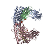
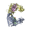
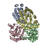
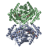
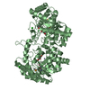
BIOMOLECULE: 1, 2, 3 SEE REMARK 350 FOR THE AUTHOR PROVIDED AND PROGRAM GENERATED ASSEMBLY ... BIOMOLECULE: 1, 2, 3 SEE REMARK 350 FOR THE AUTHOR PROVIDED AND PROGRAM GENERATED ASSEMBLY INFORMATION FOR THE STRUCTURE IN THIS ENTRY. THE REMARK MAY ALSO PROVIDE INFORMATION ON BURIED SURFACE AREA. THIS ENTRY CONTAINS THE CRYSTALLOGRAPHIC ASYMMETRIC UNIT WHICH CONSISTS OF 2 CHAINS. AUTHORS STATE THAT THE EBI/PISA ANALYSIS SUPPORTS THE ASSIGNMENT OF A TETRAMER AS A SIGNIFICANT OLIGOMERIZATION STATE.
Remark 999
SEQUENCE THE CONSTRUCT WAS EXPRESSED WITH A PURIFICATION TAG MGSDKIHHHHHHENLYFQG. THE TAG WAS ... SEQUENCE THE CONSTRUCT WAS EXPRESSED WITH A PURIFICATION TAG MGSDKIHHHHHHENLYFQG. THE TAG WAS REMOVED WITH TEV PROTEASE LEAVING ONLY A GLYCINE (0) FOLLOWED BY THE TARGET SEQUENCE.
Monochromator: Single crystal Si(111) bent (horizontal focusing) Protocol: MAD / Monochromatic (M) / Laue (L): M / Scattering type: x-ray
Radiation wavelength
ID
Wavelength (Å)
Relative weight
1
0.91837
1
2
0.97939
1
Reflection
Resolution: 1.78→42.108 Å / Num. obs: 71702 / % possible obs: 97.5 % / Biso Wilson estimate: 25.55 Å2 / Rmerge(I) obs: 0.108 / Net I/σ(I): 14.47
Reflection shell
Resolution (Å)
Rmerge(I) obs
Mean I/σ(I) obs
Num. measured obs
Num. unique all
Diffraction-ID
% possible all
1.78-1.84
0.684
2.3
26254
5491
1
81
1.84-1.92
0.621
3.1
49074
7575
1
97.4
1.92-2
0.51
4.2
46544
6464
1
98.4
2-2.11
0.389
5.5
54084
7465
1
99.3
2.11-2.24
0.298
7.1
51409
7042
1
99.4
2.24-2.41
0.223
9.4
51868
7117
1
99.5
2.41-2.66
0.167
12.2
54360
7450
1
99.7
2.66-3.04
0.107
18.2
52396
7223
1
99.8
3.04-42.108
0.054
31.4
53204
7419
1
99.9
-
Phasing
Phasing
Method: MAD
-
Processing
Software
Name
Version
Classification
NB
REFMAC
5.2.0019
refinement
PHENIX
refinement
SHELX
phasing
MolProbity
3beta29
modelbuilding
XSCALE
datascaling
PDB_EXTRACT
2
dataextraction
MAR345
CCD
datacollection
XDS
datareduction
autoSHARP
phasing
Refinement
Method to determine structure: MAD / Resolution: 1.78→42.108 Å / Cor.coef. Fo:Fc: 0.962 / Cor.coef. Fo:Fc free: 0.937 / SU B: 3.321 / SU ML: 0.098 / Cross valid method: THROUGHOUT / σ(F): 0 / ESU R: 0.12 / ESU R Free: 0.122 Stereochemistry target values: MAXIMUM LIKELIHOOD WITH PHASES Details: 1. HYDROGENS HAVE BEEN ADDED IN THE RIDING POSITIONS. 2. A MET-INHIBITION PROTOCOL WAS USED FOR SELENOMETHIONINE INCORPORATION DURING PROTEIN EXPRESSION. THE OCCUPANCY OF THE SE ATOMS IN THE ...Details: 1. HYDROGENS HAVE BEEN ADDED IN THE RIDING POSITIONS. 2. A MET-INHIBITION PROTOCOL WAS USED FOR SELENOMETHIONINE INCORPORATION DURING PROTEIN EXPRESSION. THE OCCUPANCY OF THE SE ATOMS IN THE MSE RESIDUES WAS REDUCED TO 0.75 FOR THE REDUCED SCATTERING POWER DUE TO PARTIAL S-MET INCORPORATION. 3. RESIDUES 0-5 IN CHAIN A AND 0-6 IN CHAIN B ARE DISORDERED AND NOT INCLUDED IN THE MODEL. 4. ZN IONS ARE MODELED BASED ON THE METAL EXCITATION SCAN. 5. A GLYCEROL MOLECULE FROM THE CRYO SOLUTION IS MODELED. 6. A LIGAND MOLECULE XYLOSE IS MODELED IN EACH MONOMER.
Rfactor
Num. reflection
% reflection
Selection details
Rfree
0.224
3610
5 %
RANDOM
Rwork
0.177
-
-
-
all
0.179
-
-
-
obs
0.179
71600
99.03 %
-
Solvent computation
Ion probe radii: 0.8 Å / Shrinkage radii: 0.8 Å / VDW probe radii: 1.2 Å / Solvent model: BABINET MODEL WITH MASK
Displacement parameters
Biso mean: 18.233 Å2
Baniso -1
Baniso -2
Baniso -3
1-
-0.79 Å2
0 Å2
0 Å2
2-
-
-0.79 Å2
0 Å2
3-
-
-
1.58 Å2
Refinement step
Cycle: LAST / Resolution: 1.78→42.108 Å
Protein
Nucleic acid
Ligand
Solvent
Total
Num. atoms
5256
0
29
690
5975
Refine LS restraints
Refine-ID
Type
Dev ideal
Dev ideal target
Number
X-RAY DIFFRACTION
r_bond_refined_d
0.017
0.022
5527
X-RAY DIFFRACTION
r_bond_other_d
0.004
0.02
3803
X-RAY DIFFRACTION
r_angle_refined_deg
1.711
1.968
7542
X-RAY DIFFRACTION
r_angle_other_deg
1.38
3
9303
X-RAY DIFFRACTION
r_dihedral_angle_1_deg
4.242
5
680
X-RAY DIFFRACTION
r_dihedral_angle_2_deg
34.548
24.457
267
X-RAY DIFFRACTION
r_dihedral_angle_3_deg
11.659
15
931
X-RAY DIFFRACTION
r_dihedral_angle_4_deg
13.049
15
30
X-RAY DIFFRACTION
r_chiral_restr
0.107
0.2
822
X-RAY DIFFRACTION
r_gen_planes_refined
0.007
0.02
6114
X-RAY DIFFRACTION
r_gen_planes_other
0.002
0.02
1098
X-RAY DIFFRACTION
r_nbd_refined
0.183
0.2
1065
X-RAY DIFFRACTION
r_nbd_other
0.147
0.2
3860
X-RAY DIFFRACTION
r_nbtor_refined
0.164
0.2
2720
X-RAY DIFFRACTION
r_nbtor_other
0.074
0.2
2597
X-RAY DIFFRACTION
r_xyhbond_nbd_refined
0.115
0.2
519
X-RAY DIFFRACTION
r_metal_ion_refined
0.124
0.2
1
X-RAY DIFFRACTION
r_symmetry_vdw_refined
0.095
0.2
24
X-RAY DIFFRACTION
r_symmetry_vdw_other
0.219
0.2
111
X-RAY DIFFRACTION
r_symmetry_hbond_refined
0.102
0.2
45
X-RAY DIFFRACTION
r_mcbond_it
2.289
3
3817
X-RAY DIFFRACTION
r_mcbond_other
0.563
3
1305
X-RAY DIFFRACTION
r_mcangle_it
2.825
5
5428
X-RAY DIFFRACTION
r_scbond_it
5.382
8
2487
X-RAY DIFFRACTION
r_scangle_it
7.218
11
2099
LS refinement shell
Resolution: 1.78→1.83 Å / Total num. of bins used: 20
Rfactor
Num. reflection
% reflection
Rfree
0.372
229
-
Rwork
0.308
4458
-
obs
-
4687
88.47 %
+
About Yorodumi
-
News
-
Feb 9, 2022. New format data for meta-information of EMDB entries
New format data for meta-information of EMDB entries
Version 3 of the EMDB header file is now the official format.
The previous official version 1.9 will be removed from the archive.
In the structure databanks used in Yorodumi, some data are registered as the other names, "COVID-19 virus" and "2019-nCoV". Here are the details of the virus and the list of structure data.
Jan 31, 2019. EMDB accession codes are about to change! (news from PDBe EMDB page)
EMDB accession codes are about to change! (news from PDBe EMDB page)
The allocation of 4 digits for EMDB accession codes will soon come to an end. Whilst these codes will remain in use, new EMDB accession codes will include an additional digit and will expand incrementally as the available range of codes is exhausted. The current 4-digit format prefixed with “EMD-” (i.e. EMD-XXXX) will advance to a 5-digit format (i.e. EMD-XXXXX), and so on. It is currently estimated that the 4-digit codes will be depleted around Spring 2019, at which point the 5-digit format will come into force.
The EM Navigator/Yorodumi systems omit the EMD- prefix.
Related info.:Q: What is EMD? / ID/Accession-code notation in Yorodumi/EM Navigator
Yorodumi is a browser for structure data from EMDB, PDB, SASBDB, etc.
This page is also the successor to EM Navigator detail page, and also detail information page/front-end page for Omokage search.
The word "yorodu" (or yorozu) is an old Japanese word meaning "ten thousand". "mi" (miru) is to see.
Related info.:EMDB / PDB / SASBDB / Comparison of 3 databanks / Yorodumi Search / Aug 31, 2016. New EM Navigator & Yorodumi / Yorodumi Papers / Jmol/JSmol / Function and homology information / Changes in new EM Navigator and Yorodumi
 Movie
Movie Controller

 Yorodumi
 Open data
 Basic information
 Components
 Keywords
 Function and homology information
 Anabaena variabilis ATCC 29413 (bacteria)
 X-RAY DIFFRACTION /
  Authors
 Citation
 Structure visualization
 Downloads & links
 Other downloads


 PDBj
PDBj
 Assembly
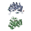

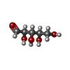
 Type: D-saccharide / Mass: 150.130 Da / Num. of mol.: 2


 Mass: 65.409 Da / Num. of mol.: 2 / Source method: obtained synthetically / Formula: Zn
 Mass: 35.453 Da / Num. of mol.: 5 / Source method: obtained synthetically / Formula: Cl
 Mass: 92.094 Da / Num. of mol.: 1 / Source method: obtained synthetically / Formula: C3H8O3
 Sample preparation
 / Beamline: BL11-1 / Wavelength: 0.91837, 0.97939
 Processing