 Movie
Movie Controller

[English] 日本語
 Yorodumi
- PDB-2qvg: The crystal structure of a two-component response regulator from ... -
+ Open data

- Basic information
| Entry | Database: PDB / ID: 2qvg | ||||||
|---|---|---|---|---|---|---|---|
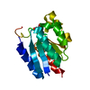
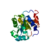
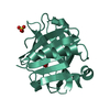
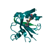
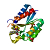
| Title | The crystal structure of a two-component response regulator from Legionella pneumophila | ||||||
 Components Components | Two component response regulator | ||||||
 Keywords Keywords | TRANSFERASE / 11016o / NYSGXRC / two component response regulator / PSI-2 / Structural Genomics / Protein Structure Initiative / New York SGX Research Center for Structural Genomics | ||||||
| Function / homology |  Function and homology information Function and homology informationTransferases; Transferring phosphorus-containing groups; Phosphotransferases with a nitrogenous group as acceptor / phosphorelay signal transduction system / transferase activity Similarity search - Function | ||||||
| Biological species |  Legionella pneumophila subsp. pneumophila str. Philadelphia 1 (bacteria) Legionella pneumophila subsp. pneumophila str. Philadelphia 1 (bacteria) | ||||||
| Method |  X-RAY DIFFRACTION / SYNCHROTRON / SAD / Resolution: 1.5 Å X-RAY DIFFRACTION / SYNCHROTRON / SAD / Resolution: 1.5 Å | ||||||
 Authors Authors | Tyagi, R. / Eswaramoorthy, S. / Burley, S.K. / Swaminathan, S. / New York SGX Research Center for Structural Genomics (NYSGXRC) | ||||||
 Citation Citation | Journal: To be Published Title: The crystal structure of a two-component response regulator from Legionella pneumophila. Authors: Tyagi, R. / Eswaramoorthy, S. / Burley, S.K. / Swaminathan, S. | ||||||
| History |
|
- Structure visualization
| Structure viewer | Molecule: MolmilJmol/JSmol |
|---|
- Downloads & links
-Download
| PDBx/mmCIF format | 2qvg.cif.gz | 42.8 KB | Display | PDBx/mmCIF format |
|---|---|---|---|---|
| PDB format | pdb2qvg.ent.gz | 29 KB | Display | PDB format |
| PDBx/mmJSON format | 2qvg.json.gz | Tree view | PDBx/mmJSON format | |
| Others |  Other downloads Other downloads |
-Validation report
| Summary document | 2qvg_validation.pdf.gz | 419.6 KB | Display | wwPDB validaton report |
|---|---|---|---|---|
| Full document | 2qvg_full_validation.pdf.gz | 423.6 KB | Display | |
| Data in XML | 2qvg_validation.xml.gz | 9.4 KB | Display | |
| Data in CIF | 2qvg_validation.cif.gz | 13.1 KB | Display | |
| Arichive directory | https://data.pdbj.org/pub/pdb/validation_reports/qv/2qvgftp://data.pdbj.org/pub/pdb/validation_reports/qv/2qvg | HTTPS FTP |
-Related structure data
| Similar structure data | |
|---|---|
| Other databases |


-Links
 PDBj
PDBj

- Assembly
| Deposited unit | 
| ||||||||
|---|---|---|---|---|---|---|---|---|---|
| 1 |
| ||||||||
| Unit cell |
| ||||||||
| Components on special symmetry positions |
|
-Components
| #1: Protein | Mass: 16692.590 Da / Num. of mol.: 1 Source method: isolated from a genetically manipulated source Source: (gene. exp.) Legionella pneumophila subsp. pneumophila str. Philadelphia 1 (bacteria)Species: Legionella pneumophila / Strain: Philadelphia 1, DSM 7513 / Gene: lpg2457 / Plasmid: pSGX3-BC / Species (production host): Escherichia coli / Production host: References: UniProt: Q5ZSR0, Transferases; Transferring phosphorus-containing groups; Phosphotransferases with a nitrogenous group as acceptor |
|---|---|
| #2: Water | ChemComp-HOH /  Mass: 18.015 Da / Num. of mol.: 171 / Source method: isolated from a natural source / Formula: H2O Mass: 18.015 Da / Num. of mol.: 171 / Source method: isolated from a natural source / Formula: H2O |
| Has protein modification | Y |
-Experimental details
-Experiment
| Experiment | Method: X-RAY DIFFRACTION / Number of used crystals: 1 |
|---|
- Sample preparation
| Crystal | Density Matthews: 2.38 Å3/Da / Density % sol: 48.34 % |
|---|---|
| Crystal grow | Temperature: 294 K / Method: vapor diffusion, sitting drop / pH: 5.5 Details: 0.2M Lithium sulfate, 0.1M Bis-Tris pH 5.5, 25% PEG 3350, VAPOR DIFFUSION, SITTING DROP, temperature 294K |
-Data collection
| Diffraction | Mean temperature: 100 K |
|---|---|
| Diffraction source | Source: SYNCHROTRON / Site: NSLS  / Beamline: X29A / Wavelength: 0.9791 Å / Beamline: X29A / Wavelength: 0.9791 Å |
| Detector | Type: ADSC QUANTUM 315 / Detector: CCD / Date: Aug 3, 2007 / Details: Mirrors |
| Radiation | Monochromator: Si(III) / Protocol: SINGLE WAVELENGTH / Monochromatic (M) / Laue (L): M / Scattering type: x-ray |
| Radiation wavelength | Wavelength: 0.9791 Å / Relative weight: 1 |
| Reflection | Resolution: 1.5→50 Å / Num. all: 44110 / Num. obs: 44110 / % possible obs: 89.1 % / Redundancy: 9.5 % / Biso Wilson estimate: 16.4 Å2 / Rmerge(I) obs: 0.047 / Net I/σ(I): 21.7 |
| Reflection shell | Resolution: 1.5→1.55 Å / Redundancy: 2.9 % / Rmerge(I) obs: 0.156 / Mean I/σ(I) obs: 7 / Num. unique all: 2265 / % possible all: 45.6 |
- Processing
| Software |
| |||||||||||||||||||||||||
|---|---|---|---|---|---|---|---|---|---|---|---|---|---|---|---|---|---|---|---|---|---|---|---|---|---|---|
| Refinement | Method to determine structure: SAD / Resolution: 1.5→40.2 Å / Rfactor Rfree error: 0.01 / Data cutoff high absF: 1225649.46 / Data cutoff low absF: 0 / Isotropic thermal model: RESTRAINED / Cross valid method: THROUGHOUT / σ(F): 0 / Stereochemistry target values: Engh & Huber Details: The Bijvoet differences were used for phasing. Residues listed as missing in Remark 465 are due to lack of electron density. Residues with missing atoms listed in Remark 470 are due to lack ...Details: The Bijvoet differences were used for phasing. Residues listed as missing in Remark 465 are due to lack of electron density. Residues with missing atoms listed in Remark 470 are due to lack of electron density for side chains and modeled as alanines.
| |||||||||||||||||||||||||
| Solvent computation | Solvent model: FLAT MODEL / Bsol: 45.9937 Å2 / ksol: 0.399418 e/Å3 | |||||||||||||||||||||||||
| Displacement parameters | Biso mean: 18.8 Å2
| |||||||||||||||||||||||||
| Refine analyze |
| |||||||||||||||||||||||||
| Refinement step | Cycle: LAST / Resolution: 1.5→40.2 Å
| |||||||||||||||||||||||||
| Refine LS restraints |
| |||||||||||||||||||||||||
| LS refinement shell | Resolution: 1.5→1.59 Å / Rfactor Rfree error: 0.03 / Total num. of bins used: 6
| |||||||||||||||||||||||||
| Xplor file |
|
