 Movie
Movie Controller

[English] 日本語
 Yorodumi
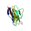
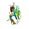
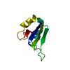
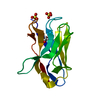
- PDB-2qsb: Crystal structure of a protein from uncharacterized family UPF014... -
+ Open data

- Basic information
| Entry | Database: PDB / ID: 2qsb | ||||||
|---|---|---|---|---|---|---|---|
| Title | Crystal structure of a protein from uncharacterized family UPF0147 from Thermoplasma acidophilum | ||||||
 Components Components | UPF0147 protein Ta0600 | ||||||
 Keywords Keywords | STRUCTURAL GENOMICS / UNKNOWN FUNCTION / four-helix bundle / Thermoplasma acidophilum / PSI-2 / Protein Structure Initiative / Midwest Center for Structural Genomics / MCSG | ||||||
| Function / homology | Uncharacterised protein family UPF0147 / Uncharacterised protein family (UPF0147) / Ta0600-like / Ta0600-like superfamily / de novo design (two linked rop proteins) / Up-down Bundle / Mainly Alpha / UPF0147 protein Ta0600 Function and homology information Function and homology information | ||||||
| Biological species |   Thermoplasma acidophilum DSM 1728 (acidophilic) Thermoplasma acidophilum DSM 1728 (acidophilic) | ||||||
| Method |  X-RAY DIFFRACTION / SYNCHROTRON / MAD / Resolution: 1.3 Å X-RAY DIFFRACTION / SYNCHROTRON / MAD / Resolution: 1.3 Å | ||||||
 Authors Authors | Cuff, M.E. / Duggan, E. / Gu, M. / Joachimiak, A. / Midwest Center for Structural Genomics (MCSG) | ||||||
 Citation Citation | Journal: TO BE PUBLISHED Title: Structure of a protein from uncharacterized family UPF0147 from Thermoplasma acidophilum. Authors: Cuff, M.E. / Duggan, E. / Gu, M. / Joachimiak, A. | ||||||
| History |
| ||||||
| Remark 300 | BIOMOLECULE: 1 SEE REMARK 350 FOR THE PROGRAM GENERATED ASSEMBLY INFORMATION FOR THE STRUCTURE IN ... BIOMOLECULE: 1 SEE REMARK 350 FOR THE PROGRAM GENERATED ASSEMBLY INFORMATION FOR THE STRUCTURE IN THIS ENTRY. AUTHORS STATE THAT THE BIOLOGICAL UNIT OF THIS POLYPEPTIDE IS UNKNOWN. |
- Structure visualization
| Structure viewer | Molecule: MolmilJmol/JSmol |
|---|
- Downloads & links
-Download
| PDBx/mmCIF format | 2qsb.cif.gz | 61.4 KB | Display | PDBx/mmCIF format |
|---|---|---|---|---|
| PDB format | pdb2qsb.ent.gz | 44.7 KB | Display | PDB format |
| PDBx/mmJSON format | 2qsb.json.gz | Tree view | PDBx/mmJSON format | |
| Others |  Other downloads Other downloads |
-Validation report
| Arichive directory | https://data.pdbj.org/pub/pdb/validation_reports/qs/2qsbftp://data.pdbj.org/pub/pdb/validation_reports/qs/2qsb | HTTPS FTP |
|---|
-Related structure data
| Similar structure data | |
|---|---|
| Other databases |
-Links
 PDBj
PDBj- Assembly
| Deposited unit | 
| ||||||||
|---|---|---|---|---|---|---|---|---|---|
| 1 |
| ||||||||
| Unit cell |
|
-Components
| #1: Protein | Mass: 10163.872 Da / Num. of mol.: 1 Source method: isolated from a genetically manipulated source Source: (gene. exp.) Thermoplasma acidophilum DSM 1728 (acidophilic)Species: Thermoplasma acidophilum / Strain: DSM 1728, IFO 15155, JCM 9062, AMRC-C165 / Gene: CAC11739, Ta0600 / Plasmid: pMCSG7 / Species (production host): Escherichia coli / Production host:  |
|---|---|
| #2: Chemical | ChemComp-SO4 /   Mass: 96.063 Da / Num. of mol.: 1 / Source method: obtained synthetically / Formula: SO4 Mass: 96.063 Da / Num. of mol.: 1 / Source method: obtained synthetically / Formula: SO4 |
| #3: Water | ChemComp-HOH /  Mass: 18.015 Da / Num. of mol.: 128 / Source method: isolated from a natural source / Formula: H2O Mass: 18.015 Da / Num. of mol.: 128 / Source method: isolated from a natural source / Formula: H2O |
| Has protein modification | Y |
-Experimental details
-Experiment
| Experiment | Method: X-RAY DIFFRACTION / Number of used crystals: 1 |
|---|
- Sample preparation
| Crystal | Density Matthews: 2.15 Å3/Da / Density % sol: 42.9 % |
|---|---|
| Crystal grow | Temperature: 291 K / Method: vapor diffusion, sitting drop / pH: 7 Details: 2M Ammonium sulfate, 0.1M Tri-HCl pH 7.0, 0.2M Lithium sulfate, VAPOR DIFFUSION, SITTING DROP, temperature 291K |
-Data collection
| Diffraction | Mean temperature: 100 K | |||||||||
|---|---|---|---|---|---|---|---|---|---|---|
| Diffraction source | Source: SYNCHROTRON / Site: APS  / Beamline: 19-ID / Wavelength: 0.97931, 0.97949 / Beamline: 19-ID / Wavelength: 0.97931, 0.97949 | |||||||||
| Detector | Type: ADSC QUANTUM 315 / Detector: CCD / Date: Dec 3, 2006 | |||||||||
| Radiation | Monochromator: SAGITALLY FOCUSED Si(111) / Protocol: MAD / Monochromatic (M) / Laue (L): M / Scattering type: x-ray | |||||||||
| Radiation wavelength |
| |||||||||
| Reflection | Resolution: 1.25→50 Å / Num. all: 23474 / Num. obs: 23474 / % possible obs: 98.2 % / Observed criterion σ(I): -3 / Redundancy: 11.1 % / Biso Wilson estimate: 11.8 Å2 / Rmerge(I) obs: 0.099 / Χ2: 3.714 / Net I/σ(I): 9.4 | |||||||||
| Reflection shell | Resolution: 1.25→1.35 Å / Redundancy: 8.9 % / Rmerge(I) obs: 0.37 / Mean I/σ(I) obs: 3.5 / Num. unique all: 1515 / Χ2: 0.908 / % possible all: 97.3 |
-Phasing
| Phasing | Method: MAD | ||||||||||||||||||||||||||||||||||||||||||||||||||||||||||||||||||||||||||||||||||||||||||||||||||||||||||||||||||||||||||||||||||||||||||||||||||||||||||||||||||||||||||
|---|---|---|---|---|---|---|---|---|---|---|---|---|---|---|---|---|---|---|---|---|---|---|---|---|---|---|---|---|---|---|---|---|---|---|---|---|---|---|---|---|---|---|---|---|---|---|---|---|---|---|---|---|---|---|---|---|---|---|---|---|---|---|---|---|---|---|---|---|---|---|---|---|---|---|---|---|---|---|---|---|---|---|---|---|---|---|---|---|---|---|---|---|---|---|---|---|---|---|---|---|---|---|---|---|---|---|---|---|---|---|---|---|---|---|---|---|---|---|---|---|---|---|---|---|---|---|---|---|---|---|---|---|---|---|---|---|---|---|---|---|---|---|---|---|---|---|---|---|---|---|---|---|---|---|---|---|---|---|---|---|---|---|---|---|---|---|---|---|---|---|---|
| Phasing MAD | D res high: 1.3 Å / D res low: 43.81 Å / FOM : 0.602 / FOM acentric: 0.608 / FOM centric: 0.398 / Reflection: 21124 / Reflection acentric: 20467 / Reflection centric: 657 | ||||||||||||||||||||||||||||||||||||||||||||||||||||||||||||||||||||||||||||||||||||||||||||||||||||||||||||||||||||||||||||||||||||||||||||||||||||||||||||||||||||||||||
| Phasing MAD set | Highest resolution: 1.3 Å / Lowest resolution: 43.81 Å
| ||||||||||||||||||||||||||||||||||||||||||||||||||||||||||||||||||||||||||||||||||||||||||||||||||||||||||||||||||||||||||||||||||||||||||||||||||||||||||||||||||||||||||
| Phasing MAD set shell |
| ||||||||||||||||||||||||||||||||||||||||||||||||||||||||||||||||||||||||||||||||||||||||||||||||||||||||||||||||||||||||||||||||||||||||||||||||||||||||||||||||||||||||||
| Phasing MAD set site |
| ||||||||||||||||||||||||||||||||||||||||||||||||||||||||||||||||||||||||||||||||||||||||||||||||||||||||||||||||||||||||||||||||||||||||||||||||||||||||||||||||||||||||||
| Phasing MAD shell |
| ||||||||||||||||||||||||||||||||||||||||||||||||||||||||||||||||||||||||||||||||||||||||||||||||||||||||||||||||||||||||||||||||||||||||||||||||||||||||||||||||||||||||||
| Phasing dm | Method: Solvent flattening and Histogram matching / Reflection: 23416 | ||||||||||||||||||||||||||||||||||||||||||||||||||||||||||||||||||||||||||||||||||||||||||||||||||||||||||||||||||||||||||||||||||||||||||||||||||||||||||||||||||||||||||
| Phasing dm shell |
|
- Processing
| Software |
| |||||||||||||||||||||||||||||||||||||||||||||||||||||||||||||||||||||||||||||||||||||||||||||||||||||||||
|---|---|---|---|---|---|---|---|---|---|---|---|---|---|---|---|---|---|---|---|---|---|---|---|---|---|---|---|---|---|---|---|---|---|---|---|---|---|---|---|---|---|---|---|---|---|---|---|---|---|---|---|---|---|---|---|---|---|---|---|---|---|---|---|---|---|---|---|---|---|---|---|---|---|---|---|---|---|---|---|---|---|---|---|---|---|---|---|---|---|---|---|---|---|---|---|---|---|---|---|---|---|---|---|---|---|---|
| Refinement | Method to determine structure: MAD / Resolution: 1.3→24.55 Å / Cor.coef. Fo:Fc: 0.973 / Cor.coef. Fo:Fc free: 0.965 / SU B: 1.346 / SU ML: 0.027 / Cross valid method: THROUGHOUT / σ(F): 0 / ESU R: 0.059 / ESU R Free: 0.053 Stereochemistry target values: MAXIMUM LIKELIHOOD WITH PHASES Details: HYDROGENS HAVE BEEN ADDED IN THE RIDING POSITIONS
| |||||||||||||||||||||||||||||||||||||||||||||||||||||||||||||||||||||||||||||||||||||||||||||||||||||||||
| Solvent computation | Ion probe radii: 0.8 Å / Shrinkage radii: 0.8 Å / VDW probe radii: 1.2 Å / Solvent model: MASK | |||||||||||||||||||||||||||||||||||||||||||||||||||||||||||||||||||||||||||||||||||||||||||||||||||||||||
| Displacement parameters | Biso mean: 16.124 Å2
| |||||||||||||||||||||||||||||||||||||||||||||||||||||||||||||||||||||||||||||||||||||||||||||||||||||||||
| Refinement step | Cycle: LAST / Resolution: 1.3→24.55 Å
| |||||||||||||||||||||||||||||||||||||||||||||||||||||||||||||||||||||||||||||||||||||||||||||||||||||||||
| Refine LS restraints |
| |||||||||||||||||||||||||||||||||||||||||||||||||||||||||||||||||||||||||||||||||||||||||||||||||||||||||
| LS refinement shell | Resolution: 1.3→1.334 Å / Total num. of bins used: 20
|
