| Entry | Database: PDB / ID: 2qr5
|
|---|
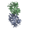
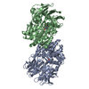
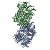
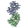
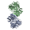
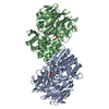
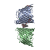
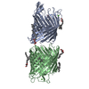
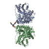
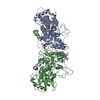
| Title | Aeropyrum pernix acylaminoacyl peptidase, H367A mutant |
|---|
 Components Components | Acylamino-acid-releasing enzyme |
|---|
 Keywords Keywords | HYDROLASE / acylaminoacyl peptidase / thermophilic enzyme / oxyanion binding site / catalytic activity |
|---|
| Function / homology |  Function and homology information Function and homology information
acylaminoacyl-peptidase / omega peptidase activity / serine-type endopeptidase activity / proteolysis / cytoplasmSimilarity search - Function Peptidase/esterase 'gauge' domain / : / Acylamino-acid-releasing enzyme-like, N-terminal domain / Peptidase S9, prolyl oligopeptidase, catalytic domain / Prolyl oligopeptidase family / 7 Propeller / Methylamine Dehydrogenase; Chain H / Alpha/Beta hydrolase fold, catalytic domain / Alpha/Beta hydrolase fold / Rossmann fold ...Peptidase/esterase 'gauge' domain / : / Acylamino-acid-releasing enzyme-like, N-terminal domain / Peptidase S9, prolyl oligopeptidase, catalytic domain / Prolyl oligopeptidase family / 7 Propeller / Methylamine Dehydrogenase; Chain H / Alpha/Beta hydrolase fold, catalytic domain / Alpha/Beta hydrolase fold / Rossmann fold / 3-Layer(aba) Sandwich / Mainly Beta / Alpha BetaSimilarity search - Domain/homology |
|---|
| Biological species |    Aeropyrum pernix (archaea) Aeropyrum pernix (archaea) |
|---|
| Method |  X-RAY DIFFRACTION / MOLECULAR REPLACEMENT / Resolution: 2.2 Å X-RAY DIFFRACTION / MOLECULAR REPLACEMENT / Resolution: 2.2 Å |
|---|
 Authors Authors | Harmat, V. / Pallo, A. / Kiss, A.L. / Polgar, L. |
|---|
 Citation Citation | Journal: J.Struct.Biol. / Year: 2008
Title: Structural and kinetic contributions of the oxyanion binding site to the catalytic activity of acylaminoacyl peptidase
Authors: Kiss, A.L. / Pallo, A. / Naray-Szabo, G. / Harmat, V. / Polgar, L. |
|---|
| History | | Deposition | Jul 27, 2007 | Deposition site: RCSB / Processing site: RCSB |
|---|
| Revision 1.0 | May 20, 2008 | Provider: repository / Type: Initial release |
|---|
| Revision 1.1 | Jul 13, 2011 | Group: Advisory / Version format compliance |
|---|
| Revision 1.2 | Oct 25, 2017 | Group: Refinement description / Category: software / Item: _software.name |
|---|
| Revision 1.3 | Oct 20, 2021 | Group: Database references / Category: database_2 / struct_ref_seq_dif
Item: _database_2.pdbx_DOI / _database_2.pdbx_database_accession / _struct_ref_seq_dif.details |
|---|
| Revision 1.4 | Aug 30, 2023 | Group: Data collection / Refinement description
Category: chem_comp_atom / chem_comp_bond ...chem_comp_atom / chem_comp_bond / pdbx_initial_refinement_model / struct_ncs_dom_lim
Item: _struct_ncs_dom_lim.beg_auth_comp_id / _struct_ncs_dom_lim.end_auth_comp_id |
|---|
|
|---|
 Movie
Movie Controller


 Open data
 Basic information
 Structure visualization
 Downloads & links
 Other downloads
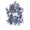

 PDBj
PDBj
 Assembly