- PDB-2qjg: M. jannaschii ADH synthase complexed with F1,6P -
+
Open data
ID or keywords:
Loading...
-
Basic information
Entry
Database: PDB / ID: 2qjg
Title
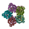
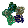
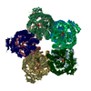
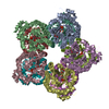
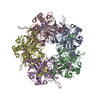
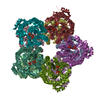
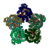
M. jannaschii ADH synthase complexed with F1,6P
Components
Putative aldolase MJ0400
Keywords
LYASE / beta-alpha barrel
Function / homology
Function and homology information
2-amino-3,7-dideoxy-D-threo-hept-6-ulosonate synthase / transketolase or transaldolase activity / fructose-bisphosphate aldolase activity / hydro-lyase activity / aromatic amino acid biosynthetic process / acyltransferase activity, transferring groups other than amino-acyl groups / amino acid biosynthetic process Similarity search - Function
2-amino-3,7-dideoxy-D-threo-hept-6-ulosonate synthase / Aldolase FbaB-like, archaeal-type / : / DeoC/LacD family aldolase / DeoC/FbaB/LacD aldolase / DeoC/LacD family aldolase / Aldolase class I / Aldolase-type TIM barrel / TIM Barrel / Alpha-Beta Barrel / Alpha Beta Similarity search - Domain/homology
In the structure databanks used in Yorodumi, some data are registered as the other names, "COVID-19 virus" and "2019-nCoV". Here are the details of the virus and the list of structure data.
Jan 31, 2019. EMDB accession codes are about to change! (news from PDBe EMDB page)
EMDB accession codes are about to change! (news from PDBe EMDB page)
The allocation of 4 digits for EMDB accession codes will soon come to an end. Whilst these codes will remain in use, new EMDB accession codes will include an additional digit and will expand incrementally as the available range of codes is exhausted. The current 4-digit format prefixed with “EMD-” (i.e. EMD-XXXX) will advance to a 5-digit format (i.e. EMD-XXXXX), and so on. It is currently estimated that the 4-digit codes will be depleted around Spring 2019, at which point the 5-digit format will come into force.
The EM Navigator/Yorodumi systems omit the EMD- prefix.
Related info.:Q: What is EMD? / ID/Accession-code notation in Yorodumi/EM Navigator
Yorodumi is a browser for structure data from EMDB, PDB, SASBDB, etc.
This page is also the successor to EM Navigator detail page, and also detail information page/front-end page for Omokage search.
The word "yorodu" (or yorozu) is an old Japanese word meaning "ten thousand". "mi" (miru) is to see.
Related info.:EMDB / PDB / SASBDB / Comparison of 3 databanks / Yorodumi Search / Aug 31, 2016. New EM Navigator & Yorodumi / Yorodumi Papers / Jmol/JSmol / Function and homology information / Changes in new EM Navigator and Yorodumi
 Movie
Movie Controller


 Open data
 Basic information
 Components
 Keywords
 Function and homology information
 Methanocaldococcus jannaschii (archaea)
 X-RAY DIFFRACTION /
  Authors
 Citation
 Structure visualization
 Downloads & links
 Other downloads


 PDBj
PDBj
 Assembly
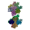

 Mass: 342.132 Da / Num. of mol.: 20 / Source method: obtained synthetically / Formula: C6H16O12P2
 Mass: 18.015 Da / Num. of mol.: 618 / Source method: isolated from a natural source / Formula: H2O
 Sample preparation
 / Beamline: 24-ID-C / Wavelength: 0.9795 Å
 Processing