- PDB-2qhd: Crystal structure of ecarpholin S (ser49-PLA2) complexed with fat... -
+
Open data
ID or keywords:
Loading...
-
Basic information
Entry
Database: PDB / ID: 2qhd
Title
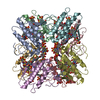
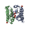
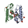
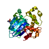
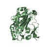
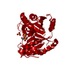
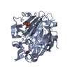
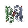
Crystal structure of ecarpholin S (ser49-PLA2) complexed with fatty acid
Components
Phospholipase A2
Keywords
HYDROLASE / BETA SHEET / THREE HELICES / PROTEIN-LIGAND COMPLEX
Function / homology
Function and homology information
A2-type glycerophospholipase activity / arachidonate secretion / lipid catabolic process / negative regulation of T cell proliferation / phospholipid metabolic process / phospholipid binding / calcium ion binding / extracellular region Similarity search - Function
Phospholipase A2, aspartic acid active site / Phospholipase A2 aspartic acid active site. / Phospholipase A2, histidine active site / Phospholipase A2 histidine active site. / Phospholipase A2 / Phospholipase A2 domain / Phospholipase A2 / Phospholipase A2 / Phospholipase A2 domain / Phospholipase A2 ...Phospholipase A2, aspartic acid active site / Phospholipase A2 aspartic acid active site. / Phospholipase A2, histidine active site / Phospholipase A2 histidine active site. / Phospholipase A2 / Phospholipase A2 domain / Phospholipase A2 / Phospholipase A2 / Phospholipase A2 domain / Phospholipase A2 / Phospholipase A2 domain superfamily / Up-down Bundle / Mainly Alpha Similarity search - Domain/homology
In the structure databanks used in Yorodumi, some data are registered as the other names, "COVID-19 virus" and "2019-nCoV". Here are the details of the virus and the list of structure data.
Jan 31, 2019. EMDB accession codes are about to change! (news from PDBe EMDB page)
EMDB accession codes are about to change! (news from PDBe EMDB page)
The allocation of 4 digits for EMDB accession codes will soon come to an end. Whilst these codes will remain in use, new EMDB accession codes will include an additional digit and will expand incrementally as the available range of codes is exhausted. The current 4-digit format prefixed with “EMD-” (i.e. EMD-XXXX) will advance to a 5-digit format (i.e. EMD-XXXXX), and so on. It is currently estimated that the 4-digit codes will be depleted around Spring 2019, at which point the 5-digit format will come into force.
The EM Navigator/Yorodumi systems omit the EMD- prefix.
Related info.:Q: What is EMD? / ID/Accession-code notation in Yorodumi/EM Navigator
Yorodumi is a browser for structure data from EMDB, PDB, SASBDB, etc.
This page is also the successor to EM Navigator detail page, and also detail information page/front-end page for Omokage search.
The word "yorodu" (or yorozu) is an old Japanese word meaning "ten thousand". "mi" (miru) is to see.
Related info.:EMDB / PDB / SASBDB / Comparison of 3 databanks / Yorodumi Search / Aug 31, 2016. New EM Navigator & Yorodumi / Yorodumi Papers / Jmol/JSmol / Function and homology information / Changes in new EM Navigator and Yorodumi
 Movie
Movie Controller

 Yorodumi
 Open data
 Basic information
 Components
 Keywords
 Function and homology information
 Echis carinatus (saw-scaled viper)
 X-RAY DIFFRACTION /
  Authors
 Citation
 Structure visualization
 Downloads & links
 Other downloads
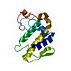


 PDBj
PDBj

 Assembly
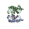

 Mass: 200.318 Da / Num. of mol.: 2 / Source method: obtained synthetically / Formula: C12H24O2
 Mass: 18.015 Da / Num. of mol.: 243 / Source method: isolated from a natural source / Formula: H2O
 Sample preparation
 / Beamline: X29A / Wavelength: 1 Å
 Processing