 Movie
Movie Controller

[English] 日本語
 Yorodumi
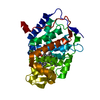
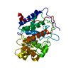
- PDB-2qha: From Structure to Function: Insights into the Catalytic Substrate... -
+ Open data

- Basic information
| Entry | Database: PDB / ID: 2qha | ||||||
|---|---|---|---|---|---|---|---|
| Title | From Structure to Function: Insights into the Catalytic Substrate Specificity and Thermostability Displayed by Bacillus subtilis mannanase BCman | ||||||
 Components Components | Beta-1,4-mannanase | ||||||
 Keywords Keywords | HYDROLASE / Beta-barrel / His1-His23-Glu336 metal-binding motif / Disulfide bond / Shallow-dish-shaped active center | ||||||
| Function / homology |  Function and homology information Function and homology informationsubstituted mannan metabolic process / mannan endo-1,4-beta-mannosidase / mannan endo-1,4-beta-mannosidase activity / extracellular region / metal ion binding Similarity search - Function | ||||||
| Biological species |  | ||||||
| Method |  X-RAY DIFFRACTION / SIRAS / Resolution: 1.45 Å X-RAY DIFFRACTION / SIRAS / Resolution: 1.45 Å | ||||||
 Authors Authors | Yan, X.X. / Liang, D.C. | ||||||
 Citation Citation | Journal: J.Mol.Biol. / Year: 2008 Title: From Structure to Function: Insights into the Catalytic Substrate Specificity and Thermostability Displayed by Bacillus subtilis Mannanase BCman Authors: Yan, X.X. / An, X.M. / Gui, L.L. / Liang, D.C. | ||||||
| History |
|
- Structure visualization
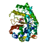
| Structure viewer | Molecule: MolmilJmol/JSmol |
|---|
- Downloads & links
-Download
| PDBx/mmCIF format | 2qha.cif.gz | 300.9 KB | Display | PDBx/mmCIF format |
|---|---|---|---|---|
| PDB format | pdb2qha.ent.gz | 244.3 KB | Display | PDB format |
| PDBx/mmJSON format | 2qha.json.gz | Tree view | PDBx/mmJSON format | |
| Others |  Other downloads Other downloads |
-Validation report
| Summary document | 2qha_validation.pdf.gz | 448 KB | Display | wwPDB validaton report |
|---|---|---|---|---|
| Full document | 2qha_full_validation.pdf.gz | 451.5 KB | Display | |
| Data in XML | 2qha_validation.xml.gz | 39.7 KB | Display | |
| Data in CIF | 2qha_validation.cif.gz | 58.3 KB | Display | |
| Arichive directory | https://data.pdbj.org/pub/pdb/validation_reports/qh/2qhaftp://data.pdbj.org/pub/pdb/validation_reports/qh/2qha | HTTPS FTP |
-Related structure data
| Similar structure data |
|---|


-Links
 PDBj
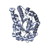
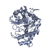
PDBj- Assembly
| Deposited unit | 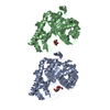
| ||||||||
|---|---|---|---|---|---|---|---|---|---|
| 1 | 
| ||||||||
| 2 | 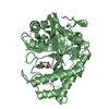
| ||||||||
| Unit cell |
|
-Components
| #1: Protein | Mass: 38963.156 Da / Num. of mol.: 2 / Fragment: UNP residues 27-362 Source method: isolated from a genetically manipulated source Source: (gene. exp.) References: UniProt: Q5PSP8, mannan endo-1,4-beta-mannosidase #2: Chemical |   Mass: 65.409 Da / Num. of mol.: 2 / Source method: obtained synthetically / Formula: Zn Mass: 65.409 Da / Num. of mol.: 2 / Source method: obtained synthetically / Formula: Zn#3: Chemical | ChemComp-GOL /   Mass: 92.094 Da / Num. of mol.: 5 / Source method: obtained synthetically / Formula: C3H8O3 Mass: 92.094 Da / Num. of mol.: 5 / Source method: obtained synthetically / Formula: C3H8O3#4: Water | ChemComp-HOH / |  Mass: 18.015 Da / Num. of mol.: 909 / Source method: isolated from a natural source / Formula: H2O Mass: 18.015 Da / Num. of mol.: 909 / Source method: isolated from a natural source / Formula: H2OHas protein modification | Y | |
|---|
-Experimental details
-Experiment
| Experiment | Method: X-RAY DIFFRACTION / Number of used crystals: 2 |
|---|
- Sample preparation
| Crystal | Density Matthews: 2.08 Å3/Da / Density % sol: 40.96 % |
|---|---|
| Crystal grow | Temperature: 290 K / Method: vapor diffusion, hanging drop / pH: 5.12 Details: 7.5% PEG4000, 80mM sodium chloride, 100mM sodium citrate (pH5.12), VAPOR DIFFUSION, HANGING DROP, temperature 290K |
-Data collection
| Diffraction | Mean temperature: 90 K |
|---|---|
| Diffraction source | Source: ROTATING ANODE / Type: RIGAKU / Wavelength: 1.5418 Å |
| Detector | Type: MAR scanner 345 mm plate / Detector: IMAGE PLATE / Date: Dec 5, 2006 |
| Radiation | Monochromator: GRAPHITE / Protocol: SINGLE WAVELENGTH / Monochromatic (M) / Laue (L): M / Scattering type: x-ray |
| Radiation wavelength | Wavelength: 1.5418 Å / Relative weight: 1 |
| Reflection | Resolution: 1.45→8 Å / Num. all: 95614 / Num. obs: 95614 / % possible obs: 90.8 % / Observed criterion σ(F): 3 / Observed criterion σ(I): 3 / Redundancy: 5.9 % / Biso Wilson estimate: 13.8 Å2 / Rmerge(I) obs: 0.034 / Net I/σ(I): 51.9 |
| Reflection shell | Resolution: 1.45→1.48 Å / Redundancy: 5.3 % / Rmerge(I) obs: 0.182 / Mean I/σ(I) obs: 11.3 / Num. unique all: 95614 / % possible all: 80.1 |
- Processing
| Software |
| ||||||||||||||||||||||||||||||||||||||||||||||||||||||||||||||||||||||||||||||||||||||||||||||||||||||||||||||
|---|---|---|---|---|---|---|---|---|---|---|---|---|---|---|---|---|---|---|---|---|---|---|---|---|---|---|---|---|---|---|---|---|---|---|---|---|---|---|---|---|---|---|---|---|---|---|---|---|---|---|---|---|---|---|---|---|---|---|---|---|---|---|---|---|---|---|---|---|---|---|---|---|---|---|---|---|---|---|---|---|---|---|---|---|---|---|---|---|---|---|---|---|---|---|---|---|---|---|---|---|---|---|---|---|---|---|---|---|---|---|---|
| Refinement | Method to determine structure: SIRAS / Resolution: 1.45→8 Å / Cor.coef. Fo:Fc: 0.968 / Cor.coef. Fo:Fc free: 0.954 / SU B: 2.033 / SU ML: 0.037 / Cross valid method: THROUGHOUT / σ(F): 3 / ESU R: 0.098 / ESU R Free: 0.074 / Stereochemistry target values: MAXIMUM LIKELIHOOD / Details: HYDROGENS HAVE BEEN ADDED IN THE RIDING POSITIONS
| ||||||||||||||||||||||||||||||||||||||||||||||||||||||||||||||||||||||||||||||||||||||||||||||||||||||||||||||
| Solvent computation | Ion probe radii: 0.8 Å / Shrinkage radii: 0.8 Å / VDW probe radii: 1.4 Å / Solvent model: MASK | ||||||||||||||||||||||||||||||||||||||||||||||||||||||||||||||||||||||||||||||||||||||||||||||||||||||||||||||
| Displacement parameters | Biso mean: 13.881 Å2
| ||||||||||||||||||||||||||||||||||||||||||||||||||||||||||||||||||||||||||||||||||||||||||||||||||||||||||||||
| Refinement step | Cycle: LAST / Resolution: 1.45→8 Å
| ||||||||||||||||||||||||||||||||||||||||||||||||||||||||||||||||||||||||||||||||||||||||||||||||||||||||||||||
| Refine LS restraints |
| ||||||||||||||||||||||||||||||||||||||||||||||||||||||||||||||||||||||||||||||||||||||||||||||||||||||||||||||
| LS refinement shell | Resolution: 1.45→1.486 Å / Total num. of bins used: 20
|
