[English] 日本語
Yorodumi
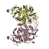
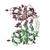
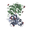
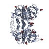
- PDB-2qfr: Crystal structure of red kidney bean purple acid phosphatase with... -

+
Open data


ID or keywords:

Loading...

-
Basic information

Entry
Database: PDB / ID: 2qfr
TitleCrystal structure of red kidney bean purple acid phosphatase with bound sulfate
ComponentsPurple acid phosphatase
KeywordsHYDROLASE / binuclear metal centre / substrate analog
Function / homology
Function and homology information


acid phosphatase / acid phosphatase activity / ferric iron binding / zinc ion binding / extracellular region
Similarity search - Function
Purple acid phosphatase-like, N-terminal / Purple acid phosphatase-like / Purple acid phosphatase, N-terminal / Iron/zinc purple acid phosphatase-like C-terminal domain / Purple acid phosphatase, metallophosphatase domain / Iron/zinc purple acid phosphatase-like protein C / Purple acid Phosphatase, N-terminal domain / Purple acid phosphatase-like, N-terminal / Metallo-dependent phosphatases / Purple Acid Phosphatase; chain A, domain 2 ...Purple acid phosphatase-like, N-terminal / Purple acid phosphatase-like / Purple acid phosphatase, N-terminal / Iron/zinc purple acid phosphatase-like C-terminal domain / Purple acid phosphatase, metallophosphatase domain / Iron/zinc purple acid phosphatase-like protein C / Purple acid Phosphatase, N-terminal domain / Purple acid phosphatase-like, N-terminal / Metallo-dependent phosphatases / Purple Acid Phosphatase; chain A, domain 2 / Calcineurin-like phosphoesterase domain, ApaH type / Calcineurin-like phosphoesterase / Metallo-dependent phosphatase-like / 4-Layer Sandwich / Immunoglobulin-like / Sandwich / Mainly Beta / Alpha Beta
Similarity search - Domain/homology
: / 2-acetamido-2-deoxy-alpha-D-glucopyranose / Fe(3+)-Zn(2+) purple acid phosphatase / Fe(3+)-Zn(2+) purple acid phosphatase
Similarity search - Component
Biological speciesPhaseolus vulgaris (French bean)
MethodX-RAY DIFFRACTION / MOLECULAR REPLACEMENT / Resolution: 2.4 Å
AuthorsGuddat, L.W. / Schenk, G. / Gahan, L.R. / Elliot, T.W. / Leung, E.
CitationJournal: Bmc Struct.Biol. / Year: 2008
Title: Crystal structures of a purple acid phosphatase, representing different steps of this enzyme's catalytic cycle.
Authors: Schenk, G. / Elliott, T.W. / Leung, E. / Carrington, L.E. / Mitic, N. / Gahan, L.R. / Guddat, L.W.
History
DepositionJun 27, 2007Deposition site: RCSB / Processing site: RCSB
Revision 1.0Oct 14, 2008Provider: repository / Type: Initial release
Revision 1.1Jul 13, 2011Group: Non-polymer description / Version format compliance
Revision 2.0Jul 29, 2020Group: Atomic model / Data collection ...Atomic model / Data collection / Derived calculations / Structure summary
Category: atom_site / chem_comp ...atom_site / chem_comp / entity / pdbx_chem_comp_identifier / pdbx_entity_nonpoly / struct_conn / struct_conn_type / struct_site / struct_site_gen
Item: _atom_site.auth_atom_id / _atom_site.label_atom_id ..._atom_site.auth_atom_id / _atom_site.label_atom_id / _chem_comp.name / _chem_comp.type / _entity.pdbx_description / _pdbx_entity_nonpoly.name / _struct_conn.conn_type_id / _struct_conn.id / _struct_conn.pdbx_dist_value / _struct_conn.pdbx_leaving_atom_flag / _struct_conn.pdbx_role / _struct_conn.ptnr1_auth_asym_id / _struct_conn.ptnr1_auth_comp_id / _struct_conn.ptnr1_auth_seq_id / _struct_conn.ptnr1_label_asym_id / _struct_conn.ptnr1_label_atom_id / _struct_conn.ptnr1_label_comp_id / _struct_conn.ptnr1_label_seq_id / _struct_conn.ptnr2_auth_asym_id / _struct_conn.ptnr2_auth_comp_id / _struct_conn.ptnr2_auth_seq_id / _struct_conn.ptnr2_label_asym_id / _struct_conn.ptnr2_label_atom_id / _struct_conn.ptnr2_label_comp_id / _struct_conn_type.id
Description: Carbohydrate remediation / Provider: repository / Type: Remediation
Revision 2.1Aug 30, 2023Group: Data collection / Database references ...Data collection / Database references / Refinement description / Structure summary
Category: chem_comp / chem_comp_atom ...chem_comp / chem_comp_atom / chem_comp_bond / database_2 / pdbx_initial_refinement_model
Item: _chem_comp.pdbx_synonyms / _database_2.pdbx_DOI / _database_2.pdbx_database_accession

-
Structure visualization

Structure viewerMolecule:
MolmilJmol/JSmol

Downloads & links

-
Assembly

Deposited unit
A: Purple acid phosphatase
B: Purple acid phosphatase
hetero molecules


Theoretical massNumber of molelcules
Total (without water)101,02118
Polymers98,6242
Non-polymers2,39616
Water7,224401
1


  • Idetical with deposited unit
  • defined by author&software
TypeNameSymmetry operationNumber
identity operation1_555x,y,z1
Buried area6230 Å2
MethodPISA
Unit cell
Length a, b, c (Å)148.030, 148.030, 160.090
Angle α, β, γ (deg.)90.000, 90.000, 90.000
Int Tables number80
Space group name H-MI41
DetailsThe biological assembly is a dimer which corresponds to the contents of the asymmetric unit.

-
Components

-
Protein , 1 types, 2 molecules AB

#1: Protein Purple acid phosphatase


Mass: 49312.117 Da / Num. of mol.: 2 / Source method: isolated from a natural source / Source: (natural) Phaseolus vulgaris (French bean)
References: UniProt: O24319, UniProt: P80366*PLUS, acid phosphatase

-
Sugars , 2 types, 8 molecules

#5: Sugar ChemComp-NDG / 2-acetamido-2-deoxy-alpha-D-glucopyranose / N-acetyl-alpha-D-glucosamine / 2-acetamido-2-deoxy-alpha-D-glucose / 2-acetamido-2-deoxy-D-glucose / 2-acetamido-2-deoxy-glucose / 2-(ACETYLAMINO)-2-DEOXY-A-D-GLUCOPYRANOSE


Type: D-saccharide, alpha linking / Mass: 221.208 Da / Num. of mol.: 2
Source method: isolated from a genetically manipulated source
Formula: C8H15NO6
IdentifierTypeProgram
DGlcpNAcaCONDENSED IUPAC CARBOHYDRATE SYMBOLGMML 1.0
N-acetyl-a-D-glucopyranosamineCOMMON NAMEGMML 1.0
a-D-GlcpNAcIUPAC CARBOHYDRATE SYMBOLPDB-CARE 1.0
GlcNAcSNFG CARBOHYDRATE SYMBOLGMML 1.0
#6: Sugar
ChemComp-NAG / 2-acetamido-2-deoxy-beta-D-glucopyranose / N-acetyl-beta-D-glucosamine / 2-acetamido-2-deoxy-beta-D-glucose / 2-acetamido-2-deoxy-D-glucose / 2-acetamido-2-deoxy-glucose / N-ACETYL-D-GLUCOSAMINE


Type: D-saccharide, beta linking / Mass: 221.208 Da / Num. of mol.: 6
Source method: isolated from a genetically manipulated source
Formula: C8H15NO6
IdentifierTypeProgram
DGlcpNAcbCONDENSED IUPAC CARBOHYDRATE SYMBOLGMML 1.0
N-acetyl-b-D-glucopyranosamineCOMMON NAMEGMML 1.0
b-D-GlcpNAcIUPAC CARBOHYDRATE SYMBOLPDB-CARE 1.0
GlcNAcSNFG CARBOHYDRATE SYMBOLGMML 1.0

-
Non-polymers , 4 types, 409 molecules

#2: Chemical ChemComp-FE / FE (III) ION


Mass: 55.845 Da / Num. of mol.: 2 / Source method: obtained synthetically / Formula: Fe
#3: Chemical ChemComp-ZN / ZINC ION


Mass: 65.409 Da / Num. of mol.: 2 / Source method: obtained synthetically / Formula: Zn
#4: Chemical
ChemComp-SO4 / SULFATE ION


Mass: 96.063 Da / Num. of mol.: 4 / Source method: obtained synthetically / Formula: SO4
#7: Water ChemComp-HOH / water


Mass: 18.015 Da / Num. of mol.: 401 / Source method: isolated from a natural source / Formula: H2O

-
Experimental details

-
Experiment

ExperimentMethod: X-RAY DIFFRACTION / Number of used crystals: 1

-
Sample preparation

CrystalDensity Matthews: 4.45 Å3/Da / Density % sol: 72.34 %
Crystal growTemperature: 291 K / Method: vapor diffusion, hanging drop / pH: 4
Details: 2M Ammonium sulfate, 0.1 M acetate pH 4.0, VAPOR DIFFUSION, HANGING DROP, temperature 291K

-
Data collection

DiffractionMean temperature: 290 K
Diffraction sourceSource: ROTATING ANODE / Type: RIGAKU FR-E+ DW / Wavelength: 1.541 Å
DetectorType: RIGAKU RAXIS IV++ / Detector: IMAGE PLATE / Date: Apr 10, 2005 / Details: Osmic
RadiationMonochromator: Mirrors / Protocol: SINGLE WAVELENGTH / Monochromatic (M) / Laue (L): M / Scattering type: x-ray
Radiation wavelengthWavelength: 1.541 Å / Relative weight: 1
ReflectionResolution: 2.4→43 Å / Num. all: 67185 / Num. obs: 55092 / % possible obs: 82.6 % / Observed criterion σ(F): 0 / Observed criterion σ(I): 0 / Redundancy: 2.76 % / Biso Wilson estimate: 38 Å2 / Rmerge(I) obs: 0.115 / Rsym value: 0.115 / Net I/σ(I): 7.1
Reflection shellResolution: 2.4→2.55 Å / Redundancy: 1.6 % / Rmerge(I) obs: 0.302 / Mean I/σ(I) obs: 2.1 / Num. unique all: 8320 / Rsym value: 0.302 / % possible all: 52

-
Processing

Software
NameVersionClassificationNB
CNSrefinement
PDB_EXTRACT2data extraction
CrystalCleardata collection
HKL-2000data reduction
HKL-2000data scaling
EPMRphasing
RefinementMethod to determine structure: MOLECULAR REPLACEMENT
Starting model: 4kbp chain b
Resolution: 2.4→43 Å / Cross valid method: throughtout / σ(F): 0 / σ(I): 0 / Stereochemistry target values: Engh & Huber
RfactorNum. reflection% reflectionSelection details
Rfree0.211 5612 8.4 %random
Rwork0.171 ---
all0.173 55092 --
obs0.173 55092 82 %-
Solvent computationBsol: 31.514 Å2
Displacement parametersBiso mean: 27.427 Å2
Baniso -1Baniso -2Baniso -3
1--1.958 Å20 Å20 Å2
2---1.958 Å20 Å2
3---3.916 Å2
Refinement stepCycle: LAST / Resolution: 2.4→43 Å
ProteinNucleic acidLigandSolventTotal
Num. atoms6974 0 138 401 7513
Refine LS restraints
Refine-IDTypeDev idealDev ideal target
X-RAY DIFFRACTIONc_bond_d0.007
X-RAY DIFFRACTIONc_angle_d1.285
X-RAY DIFFRACTIONc_mcbond_it1.0451.5
X-RAY DIFFRACTIONc_scbond_it1.9072
X-RAY DIFFRACTIONc_mcangle_it1.7332
X-RAY DIFFRACTIONc_scangle_it2.9472.5
Xplor file
Refine-IDSerial noParam fileTopol file
X-RAY DIFFRACTION1CNS_TOPPAR:protein_rep.paramCNS_TOPPAR:protein.top
X-RAY DIFFRACTION2CNS_TOPPAR:water_rep.paramCNS_TOPPAR:water.top
X-RAY DIFFRACTION3CNS_TOPPAR:ion.paramCNS_TOPPAR:ion.top
X-RAY DIFFRACTION4CNS_TOPPAR:carbohydrate.paramCNS_TOPPAR:carbohydrate.top

+
About Yorodumi

-
News

-
Feb 9, 2022. New format data for meta-information of EMDB entries

New format data for meta-information of EMDB entries

  • Version 3 of the EMDB header file is now the official format.
  • The previous official version 1.9 will be removed from the archive.

Related info.:EMDB header

External links:wwPDB to switch to version 3 of the EMDB data model

-
Aug 12, 2020. Covid-19 info

Covid-19 info

URL: https://pdbjlvh1.pdbj.org/emnavi/covid19.php

New page: Covid-19 featured information page in EM Navigator.

Related info.:Covid-19 info / Mar 5, 2020. Novel coronavirus structure data

+
Mar 5, 2020. Novel coronavirus structure data

Novel coronavirus structure data

Related info.:Yorodumi Speices / Aug 12, 2020. Covid-19 info

External links:COVID-19 featured content - PDBj / Molecule of the Month (242):Coronavirus Proteases

+
Jan 31, 2019. EMDB accession codes are about to change! (news from PDBe EMDB page)

EMDB accession codes are about to change! (news from PDBe EMDB page)

  • The allocation of 4 digits for EMDB accession codes will soon come to an end. Whilst these codes will remain in use, new EMDB accession codes will include an additional digit and will expand incrementally as the available range of codes is exhausted. The current 4-digit format prefixed with “EMD-” (i.e. EMD-XXXX) will advance to a 5-digit format (i.e. EMD-XXXXX), and so on. It is currently estimated that the 4-digit codes will be depleted around Spring 2019, at which point the 5-digit format will come into force.
  • The EM Navigator/Yorodumi systems omit the EMD- prefix.

Related info.:Q: What is EMD? / ID/Accession-code notation in Yorodumi/EM Navigator

External links:EMDB Accession Codes are Changing Soon! / Contact to PDBj

+
Jul 12, 2017. Major update of PDB

Major update of PDB

  • wwPDB released updated PDB data conforming to the new PDBx/mmCIF dictionary.
  • This is a major update changing the version number from 4 to 5, and with Remediation, in which all the entries are updated.
  • In this update, many items about electron microscopy experimental information are reorganized (e.g. em_software).
  • Now, EM Navigator and Yorodumi are based on the updated data.

External links:wwPDB Remediation / Enriched Model Files Conforming to OneDep Data Standards Now Available in the PDB FTP Archive

-
Yorodumi

Thousand views of thousand structures

  • Yorodumi is a browser for structure data from EMDB, PDB, SASBDB, etc.
  • This page is also the successor to EM Navigator detail page, and also detail information page/front-end page for Omokage search.
  • The word "yorodu" (or yorozu) is an old Japanese word meaning "ten thousand". "mi" (miru) is to see.

Related info.:EMDB / PDB / SASBDB / Comparison of 3 databanks / Yorodumi Search / Aug 31, 2016. New EM Navigator & Yorodumi / Yorodumi Papers / Jmol/JSmol / Function and homology information / Changes in new EM Navigator and Yorodumi

Read more