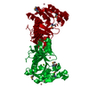
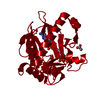
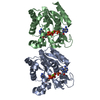
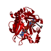
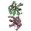
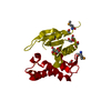
- PDB-2qec: Crystal structure of histone acetyltransferase HPA2 and related a... -
+
Open data
ID or keywords:
Loading...
-
Basic information
Entry
Database: PDB / ID: 2qec
Title
Crystal structure of histone acetyltransferase HPA2 and related acetyltransferase (NP_600742.1) from Corynebacterium glutamicum ATCC 13032 at 1.90 A resolution
Components
Histone acetyltransferase HPA2 and related acetyltransferases
Keywords
TRANSFERASE / NP_600742.1 / histone acetyltransferase HPA2 and related acetyltransferase / Acetyltransferase (GNAT) family / Structural Genomics / Joint Center for Structural Genomics / JCSG / Protein Structure Initiative / PSI-2
Mass: 18.015 Da / Num. of mol.: 151 / Source method: isolated from a natural source / Formula: H2O
Has protein modification
Y
Sequence details
1. THE CONSTRUCT WAS EXPRESSED WITH A PURIFICATION TAG MGSDKIHHHHHHENLYFQG. THE TAG WAS REMOVED ...1. THE CONSTRUCT WAS EXPRESSED WITH A PURIFICATION TAG MGSDKIHHHHHHENLYFQG. THE TAG WAS REMOVED WITH TEV PROTEASE LEAVING ONLY A GLYCINE, FOLLOWED BY THE TARGET SEQUENCE. 2. THE CONSTRUCT WAS ENGINEERED WITH THE FOLLOWING MUTATION: A29T.
-
Experimental details
-
Experiment
Experiment
Method: X-RAY DIFFRACTION / Number of used crystals: 2
-
Sample preparation
Crystal
ID
Density Matthews (Å3/Da)
Density % sol (%)
Description
1
3.67
66.5
DATA FROM A SE-MET CONTAINING CRYSTAL IN SPACEGROUP P6(2)22 WAS USED FOR THE MAD PHASING EXPERIMENTS AT 2.30 ANGSTROM RESOLUTION. THIS MAD STRUCTURE WAS USED AS A MOLECULAR REPLACEMENT MODEL TO PHASE THIS STRUCTURE AT 1.90 ANGSTROM RESOLUTION IN THE P6(5)22 SPACEGROUP.
Resolution: 1.9→27.64 Å / Num. obs: 27197 / % possible obs: 99.5 % / Redundancy: 17.62 % / Biso Wilson estimate: 32.59 Å2 / Rmerge(I) obs: 0.071 / Net I/σ(I): 17.36
Reflection shell
Resolution (Å)
Rmerge(I) obs
Mean I/σ(I) obs
Num. measured obs
Diffraction-ID
% possible all
1.9-1.97
0.905
2
29060
1,2
99.3
1.97-2.05
0.624
2.8
29134
1,2
99
2.05-2.14
0.422
4
27573
1,2
99.3
2.14-2.25
0.411
5.9
47227
1,2
99.5
2.25-2.39
0.333
8.3
57039
1,2
99.4
2.39-2.58
0.226
11.7
59495
1,2
99.6
2.58-2.84
0.144
17.3
57836
1,2
99.8
2.84-3.25
0.086
27.1
57913
1,2
99.8
3.25-4.08
0.051
42
56376
1,2
99.9
4.08-27.64
0.036
51.7
57459
1,2
99.5
-
Phasing
Phasing
Method
MAD
molecular replacement
-
Processing
Software
Name
Version
Classification
NB
MolProbity
3beta29
modelbuilding
REFMAC
5.2.0005
refinement
XSCALE
datascaling
PDB_EXTRACT
2
dataextraction
Blu-Ice
v5.0
datacollection
XDS
datareduction
MOSFLM
datareduction
SCALA
datascaling
SHELXD
phasing
autoSHARP
phasing
PHASER
phasing
Refinement
Method to determine structure: MAD, MOLECULAR REPLACEMENT / Resolution: 1.9→27.64 Å / Cor.coef. Fo:Fc: 0.96 / Cor.coef. Fo:Fc free: 0.94 / SU B: 6.388 / SU ML: 0.096 / TLS residual ADP flag: LIKELY RESIDUAL / Cross valid method: THROUGHOUT / σ(F): 0 / ESU R: 0.106 / ESU R Free: 0.116 / Stereochemistry target values: MAXIMUM LIKELIHOOD Details: 1. HYDROGENS HAVE BEEN ADDED IN THE RIDING POSITIONS. 2. A MET-INHIBITION PROTOCOL WAS USED FOR SELENOMETHIONINE INCORPORATION DURING PROTEIN EXPRESSION. THE OCCUPANCY OF THE SE ATOMS IN THE ...Details: 1. HYDROGENS HAVE BEEN ADDED IN THE RIDING POSITIONS. 2. A MET-INHIBITION PROTOCOL WAS USED FOR SELENOMETHIONINE INCORPORATION DURING PROTEIN EXPRESSION. THE OCCUPANCY OF THE SE ATOMS IN THE MSE RESIDUES WAS REDUCED TO 0.75 FOR THE REDUCED SCATTERING POWER DUE TO PARTIAL S-MET INCORPORATION. 3. ATOM RECORD CONTAINS RESIDUAL B FACTORS ONLY. 4. ETHYLENE GLYCOL WAS MODELED BASED ON CRYO CONDITIONS. 5. RESIDUES 83-94, 103, 107-115 ARE DISORDERED AND WERE NOT MODELED. 6. ASP 186 IS A RAMACHANDRAN OUTLIER AND IS LOCATED IN POOR DENSITY.
Rfactor
Num. reflection
% reflection
Selection details
Rfree
0.242
1363
5 %
RANDOM
Rwork
0.193
-
-
-
all
0.195
-
-
-
obs
0.195
27120
99.48 %
-
Solvent computation
Ion probe radii: 0.8 Å / Shrinkage radii: 0.8 Å / VDW probe radii: 1.2 Å / Solvent model: MASK
Displacement parameters
Biso mean: 41.353 Å2
Baniso -1
Baniso -2
Baniso -3
1-
1.77 Å2
0.89 Å2
0 Å2
2-
-
1.77 Å2
0 Å2
3-
-
-
-2.66 Å2
Refinement step
Cycle: LAST / Resolution: 1.9→27.64 Å
Protein
Nucleic acid
Ligand
Solvent
Total
Num. atoms
1375
0
36
151
1562
Refine LS restraints
Refine-ID
Type
Dev ideal
Dev ideal target
Number
X-RAY DIFFRACTION
r_bond_refined_d
0.016
0.022
1457
X-RAY DIFFRACTION
r_bond_other_d
0.001
0.02
1357
X-RAY DIFFRACTION
r_angle_refined_deg
1.609
1.975
1977
X-RAY DIFFRACTION
r_angle_other_deg
1.095
3
3139
X-RAY DIFFRACTION
r_dihedral_angle_1_deg
6.529
5
183
X-RAY DIFFRACTION
r_dihedral_angle_2_deg
37.366
23.158
57
X-RAY DIFFRACTION
r_dihedral_angle_3_deg
12.391
15
206
X-RAY DIFFRACTION
r_dihedral_angle_4_deg
15.97
15
9
X-RAY DIFFRACTION
r_chiral_restr
0.149
0.2
220
X-RAY DIFFRACTION
r_gen_planes_refined
0.006
0.02
1595
X-RAY DIFFRACTION
r_gen_planes_other
0.001
0.02
290
X-RAY DIFFRACTION
r_nbd_refined
0.225
0.2
286
X-RAY DIFFRACTION
r_nbd_other
0.187
0.2
1311
X-RAY DIFFRACTION
r_nbtor_refined
0.181
0.2
691
X-RAY DIFFRACTION
r_nbtor_other
0.086
0.2
834
X-RAY DIFFRACTION
r_xyhbond_nbd_refined
0.205
0.2
105
X-RAY DIFFRACTION
r_symmetry_vdw_refined
0.037
0.2
6
X-RAY DIFFRACTION
r_symmetry_vdw_other
0.195
0.2
73
X-RAY DIFFRACTION
r_symmetry_hbond_refined
0.2
0.2
13
X-RAY DIFFRACTION
r_mcbond_it
2.37
3
942
X-RAY DIFFRACTION
r_mcbond_other
0.603
3
363
X-RAY DIFFRACTION
r_mcangle_it
3.613
5
1471
X-RAY DIFFRACTION
r_scbond_it
5.4
8
601
X-RAY DIFFRACTION
r_scangle_it
7.142
11
503
LS refinement shell
Resolution: 1.9→1.949 Å / Total num. of bins used: 20
Rfactor
Num. reflection
% reflection
Rfree
0.377
87
-
Rwork
0.285
1843
-
obs
-
1930
99.13 %
Refinement TLS params.
Method: refined / Refine-ID: X-RAY DIFFRACTION
ID
L11 (°2)
L12 (°2)
L13 (°2)
L22 (°2)
L23 (°2)
L33 (°2)
S11 (Å °)
S12 (Å °)
S13 (Å °)
S21 (Å °)
S22 (Å °)
S23 (Å °)
S31 (Å °)
S32 (Å °)
S33 (Å °)
T11 (Å2)
T12 (Å2)
T13 (Å2)
T22 (Å2)
T23 (Å2)
T33 (Å2)
Origin x (Å)
Origin y (Å)
Origin z (Å)
1
1.5341
-0.1742
1.0368
0.5747
0.6867
3.9742
0.0749
-0.0492
0.0814
-0.2093
-0.246
0.2036
-0.1457
-0.3169
0.1711
-0.0148
0.0085
-0.0784
-0.2175
-0.0622
-0.1478
14.851
26.013
4.503
2
0.6385
0.4625
-0.0444
4.1358
1.7665
1.7819
0.032
0.1007
-0.0192
-0.3209
-0.0852
-0.0303
-0.2008
-0.1311
0.0532
0.0135
-0.0208
-0.0382
-0.2329
0.0157
-0.1767
21.754
11.245
0.638
3
20.1585
20.5462
7.4903
49.7283
24.3809
49.3682
-0.0307
-0.9469
-0.8857
2.894
-0.8503
1.7972
1.8854
-2.7126
0.881
0.6811
0.0576
-0.0052
0.8726
-0.093
0.7619
-1.616
22.764
2.037
Refinement TLS group
Refine-ID: X-RAY DIFFRACTION / Selection: ALL / Auth asym-ID: A / Label asym-ID: A
ID
Refine TLS-ID
Auth seq-ID
Label seq-ID
1
1
1 - 82
2 - 83
2
2
117 - 203
118 - 204
3
3
95 - 106
96 - 107
+
About Yorodumi
-
News
-
Feb 9, 2022. New format data for meta-information of EMDB entries
New format data for meta-information of EMDB entries
Version 3 of the EMDB header file is now the official format.
The previous official version 1.9 will be removed from the archive.
In the structure databanks used in Yorodumi, some data are registered as the other names, "COVID-19 virus" and "2019-nCoV". Here are the details of the virus and the list of structure data.
Jan 31, 2019. EMDB accession codes are about to change! (news from PDBe EMDB page)
EMDB accession codes are about to change! (news from PDBe EMDB page)
The allocation of 4 digits for EMDB accession codes will soon come to an end. Whilst these codes will remain in use, new EMDB accession codes will include an additional digit and will expand incrementally as the available range of codes is exhausted. The current 4-digit format prefixed with “EMD-” (i.e. EMD-XXXX) will advance to a 5-digit format (i.e. EMD-XXXXX), and so on. It is currently estimated that the 4-digit codes will be depleted around Spring 2019, at which point the 5-digit format will come into force.
The EM Navigator/Yorodumi systems omit the EMD- prefix.
Related info.:Q: What is EMD? / ID/Accession-code notation in Yorodumi/EM Navigator
Yorodumi is a browser for structure data from EMDB, PDB, SASBDB, etc.
This page is also the successor to EM Navigator detail page, and also detail information page/front-end page for Omokage search.
The word "yorodu" (or yorozu) is an old Japanese word meaning "ten thousand". "mi" (miru) is to see.
Related info.:EMDB / PDB / SASBDB / Comparison of 3 databanks / Yorodumi Search / Aug 31, 2016. New EM Navigator & Yorodumi / Yorodumi Papers / Jmol/JSmol / Function and homology information / Changes in new EM Navigator and Yorodumi
 Movie
Movie Controller

 Yorodumi
 Open data
 Basic information
 Components
 Keywords
 Function and homology information
 Corynebacterium glutamicum ATCC 13032 (bacteria)
 X-RAY DIFFRACTION /
  Authors
 Citation
 Structure visualization
 Downloads & links
 Other downloads
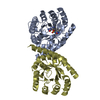


 PDBj
PDBj

 Assembly
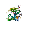

 Mass: 62.068 Da / Num. of mol.: 9 / Source method: obtained synthetically / Formula: C2H6O2
 Mass: 18.015 Da / Num. of mol.: 151 / Source method: isolated from a natural source / Formula: H2O
 Sample preparation
 Processing