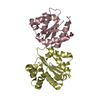
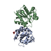
BIOMOLECULE: 1 THIS ENTRY CONTAINS THE CRYSTALLOGRAPHIC ASYMMETRIC UNIT WHICH CONSISTS OF 1 CHAINS. ... BIOMOLECULE: 1 THIS ENTRY CONTAINS THE CRYSTALLOGRAPHIC ASYMMETRIC UNIT WHICH CONSISTS OF 1 CHAINS. SEE REMARK 350 FOR INFORMATION ON GENERATING THE BIOLOGICAL MOLECULE(S). SIZE EXCLUSION CHROMATOGRAPHY WITH STATIC LIGHT SCATTERING SUPPORTS THE ASSIGNMENT OF A DIMER AS THE SIGNIFICANT OLIGOMERIZATION STATE.
Remark 999
SEQUENCE THE PROTEIN WAS EXPRESSED AND PURIFIED WITH A TAG MGSDKIHHHHHHENLYFQG FROM CONSTRUCT.
Resolution: 2→29.437 Å / Num. obs: 16338 / % possible obs: 100 % / Redundancy: 9.1 % / Biso Wilson estimate: 19.7 Å2 / Rmerge(I) obs: 0.102 / Rsym value: 0.102 / Net I/σ(I): 5.6
Reflection shell
Diffraction-ID: 1
Resolution (Å)
Redundancy (%)
Rmerge(I) obs
Mean I/σ(I) obs
Num. measured all
Num. unique all
Rsym value
% possible all
2-2.05
9.2
1.229
0.6
10741
1166
1.229
100
2.05-2.11
9.2
1.007
0.7
10525
1145
1.007
100
2.11-2.17
9.2
0.74
1
10549
1144
0.74
100
2.17-2.24
9.2
0.676
1.1
9847
1066
0.676
100
2.24-2.31
9.2
0.5
1.3
10053
1093
0.5
100
2.31-2.39
9.2
0.409
1.9
9405
1018
0.409
100
2.39-2.48
9.2
0.319
2.4
8999
980
0.319
100
2.48-2.58
9.2
0.244
3.1
9090
993
0.244
100
2.58-2.7
9.2
0.205
3.6
8427
914
0.205
100
2.7-2.83
9.1
0.168
4.3
8168
895
0.168
100
2.83-2.98
9.2
0.14
5.1
7567
824
0.14
100
2.98-3.16
9.1
0.111
6.3
7420
813
0.111
100
3.16-3.38
9.1
0.082
8
6838
753
0.082
100
3.38-3.65
9
0.067
9
6325
703
0.067
100
3.65-4
9
0.057
10.6
6044
670
0.057
100
4-4.47
8.9
0.049
11.6
5194
582
0.049
100
4.47-5.16
8.8
0.052
11.1
4714
535
0.052
100
5.16-6.32
8.7
0.057
10.7
3943
455
0.057
100
6.32-8.94
8.3
0.052
12
3059
370
0.052
100
8.94-29.44
7.4
0.043
14.3
1616
219
0.043
97.4
-
Phasing
Phasing
Method: MAD
-
Processing
Software
Name
Version
Classification
NB
REFMAC
5.2.0005
refinement
PHENIX
refinement
SOLVE
phasing
MolProbity
3beta29
modelbuilding
SCALA
datascaling
PDB_EXTRACT
3
dataextraction
MAR345
CCD
datacollection
MOSFLM
datareduction
Refinement
Method to determine structure: MAD / Resolution: 2→29.437 Å / Cor.coef. Fo:Fc: 0.963 / Cor.coef. Fo:Fc free: 0.952 / SU B: 8.222 / SU ML: 0.114 / TLS residual ADP flag: LIKELY RESIDUAL / Cross valid method: THROUGHOUT / σ(F): 0 / ESU R: 0.152 / ESU R Free: 0.143 Stereochemistry target values: MAXIMUM LIKELIHOOD WITH PHASES Details: 1. HYDROGENS HAVE BEEN ADDED IN THE RIDING POSITIONS. 2. ATOM RECORD CONTAINS RESIDUAL B FACTORS ONLY. 3. A MET-INHIBITION PROTOCOL WAS USED FOR SELENOMETHIONINE INCORPORATION DURING PROTEIN ...Details: 1. HYDROGENS HAVE BEEN ADDED IN THE RIDING POSITIONS. 2. ATOM RECORD CONTAINS RESIDUAL B FACTORS ONLY. 3. A MET-INHIBITION PROTOCOL WAS USED FOR SELENOMETHIONINE INCORPORATION DURING PROTEIN EXPRESSION. THE OCCUPANCY OF THE SE ATOMS IN THE MSE RESIDUES WAS REDUCED TO 0.75 TO ACCOUNT FOR THE REDUCED SCATTERING POWER DUE TO PARTIAL S-MET INCORPORATION. 4. ONE CHLORIDE ANION AND A CITRATE ANION (FLC) ARE MODELED IN THE STRUCTURE.
Rfactor
Num. reflection
% reflection
Selection details
Rfree
0.223
821
5 %
RANDOM
Rwork
0.185
-
-
-
obs
0.187
16309
99.94 %
-
Solvent computation
Ion probe radii: 0.8 Å / Shrinkage radii: 0.8 Å / VDW probe radii: 1.2 Å / Solvent model: MASK
Displacement parameters
Biso mean: 38.044 Å2
Baniso -1
Baniso -2
Baniso -3
1-
0.09 Å2
0.04 Å2
0 Å2
2-
-
0.09 Å2
0 Å2
3-
-
-
-0.13 Å2
Refinement step
Cycle: LAST / Resolution: 2→29.437 Å
Protein
Nucleic acid
Ligand
Solvent
Total
Num. atoms
1362
0
14
92
1468
Refine LS restraints
Refine-ID
Type
Dev ideal
Dev ideal target
Number
X-RAY DIFFRACTION
r_bond_refined_d
0.018
0.022
1472
X-RAY DIFFRACTION
r_bond_other_d
0.002
0.02
1309
X-RAY DIFFRACTION
r_angle_refined_deg
1.511
1.959
1996
X-RAY DIFFRACTION
r_angle_other_deg
0.942
3
3035
X-RAY DIFFRACTION
r_dihedral_angle_1_deg
3.647
5
177
X-RAY DIFFRACTION
r_dihedral_angle_2_deg
30.839
23.797
79
X-RAY DIFFRACTION
r_dihedral_angle_3_deg
12.379
15
259
X-RAY DIFFRACTION
r_dihedral_angle_4_deg
14.132
15
11
X-RAY DIFFRACTION
r_chiral_restr
0.12
0.2
207
X-RAY DIFFRACTION
r_gen_planes_refined
0.005
0.02
1676
X-RAY DIFFRACTION
r_gen_planes_other
0.002
0.02
334
X-RAY DIFFRACTION
r_nbd_refined
0.191
0.3
277
X-RAY DIFFRACTION
r_nbd_other
0.161
0.3
1319
X-RAY DIFFRACTION
r_nbtor_refined
0.182
0.5
738
X-RAY DIFFRACTION
r_nbtor_other
0.086
0.5
851
X-RAY DIFFRACTION
r_xyhbond_nbd_refined
0.179
0.5
116
X-RAY DIFFRACTION
r_symmetry_vdw_refined
0.114
0.3
10
X-RAY DIFFRACTION
r_symmetry_vdw_other
0.126
0.3
38
X-RAY DIFFRACTION
r_symmetry_hbond_refined
0.108
0.5
9
X-RAY DIFFRACTION
r_mcbond_it
1.618
3
901
X-RAY DIFFRACTION
r_mcbond_other
0.364
3
347
X-RAY DIFFRACTION
r_mcangle_it
2.477
5
1403
X-RAY DIFFRACTION
r_scbond_it
4.539
8
671
X-RAY DIFFRACTION
r_scangle_it
5.858
11
593
LS refinement shell
Resolution: 2→2.052 Å / Total num. of bins used: 20
Rfactor
Num. reflection
% reflection
Rfree
0.372
67
-
Rwork
0.265
1107
-
obs
-
1174
100 %
Refinement TLS params.
Method: refined / Refine-ID: X-RAY DIFFRACTION
ID
L11 (°2)
L12 (°2)
L13 (°2)
L22 (°2)
L23 (°2)
L33 (°2)
S11 (Å °)
S12 (Å °)
S13 (Å °)
S21 (Å °)
S22 (Å °)
S23 (Å °)
S31 (Å °)
S32 (Å °)
S33 (Å °)
T11 (Å2)
T12 (Å2)
T13 (Å2)
T22 (Å2)
T23 (Å2)
T33 (Å2)
Origin x (Å)
Origin y (Å)
Origin z (Å)
1
0.9278
-0.7211
-0.1321
2.8968
0.0547
2.4874
0.1624
0.1779
-0.1871
-0.2377
-0.1094
0.2435
0.183
-0.1082
-0.053
-0.0756
0.0333
-0.0416
-0.1287
-0.0355
-0.0734
-2.34
52.245
18.495
2
5.0935
-0.8331
1.6689
6.0194
-0.6101
3.125
0.5124
1.1969
-0.0784
-1.1379
-0.4377
-0.3416
0.2781
0.5622
-0.0747
0.159
0.2107
0.041
0.1475
-0.0841
-0.1803
1.719
57.258
4.47
Refinement TLS group
ID
Refine-ID
Refine TLS-ID
Auth asym-ID
Label asym-ID
Auth seq-ID
Label seq-ID
1
X-RAY DIFFRACTION
1
A
A
-2 - 99
17 - 118
2
X-RAY DIFFRACTION
2
A
A
100 - 161
119 - 180
+
About Yorodumi
-
News
-
Feb 9, 2022. New format data for meta-information of EMDB entries
New format data for meta-information of EMDB entries
Version 3 of the EMDB header file is now the official format.
The previous official version 1.9 will be removed from the archive.
In the structure databanks used in Yorodumi, some data are registered as the other names, "COVID-19 virus" and "2019-nCoV". Here are the details of the virus and the list of structure data.
Jan 31, 2019. EMDB accession codes are about to change! (news from PDBe EMDB page)
EMDB accession codes are about to change! (news from PDBe EMDB page)
The allocation of 4 digits for EMDB accession codes will soon come to an end. Whilst these codes will remain in use, new EMDB accession codes will include an additional digit and will expand incrementally as the available range of codes is exhausted. The current 4-digit format prefixed with “EMD-” (i.e. EMD-XXXX) will advance to a 5-digit format (i.e. EMD-XXXXX), and so on. It is currently estimated that the 4-digit codes will be depleted around Spring 2019, at which point the 5-digit format will come into force.
The EM Navigator/Yorodumi systems omit the EMD- prefix.
Related info.:Q: What is EMD? / ID/Accession-code notation in Yorodumi/EM Navigator
Yorodumi is a browser for structure data from EMDB, PDB, SASBDB, etc.
This page is also the successor to EM Navigator detail page, and also detail information page/front-end page for Omokage search.
The word "yorodu" (or yorozu) is an old Japanese word meaning "ten thousand". "mi" (miru) is to see.
Related info.:EMDB / PDB / SASBDB / Comparison of 3 databanks / Yorodumi Search / Aug 31, 2016. New EM Navigator & Yorodumi / Yorodumi Papers / Jmol/JSmol / Function and homology information / Changes in new EM Navigator and Yorodumi
 Movie
Movie Controller

 Yorodumi
 Open data
 Basic information
 Components
 Keywords
 Function and homology information
 Streptococcus agalactiae 2603V/R (bacteria)
 X-RAY DIFFRACTION /
  Authors
 Citation
 Structure visualization
 Downloads & links
 Other downloads
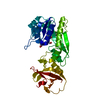


 PDBj
PDBj


 Assembly


 Mass: 35.453 Da / Num. of mol.: 1 / Source method: obtained synthetically / Formula: Cl
 Mass: 189.100 Da / Num. of mol.: 1 / Source method: obtained synthetically / Formula: C6H5O7
 Mass: 18.015 Da / Num. of mol.: 92 / Source method: isolated from a natural source / Formula: H2O
 Sample preparation
 / Beamline: BL9-2 / Wavelength: 0.91837, 0.97932
 Processing