 Movie
Movie Controller

[English] 日本語
 Yorodumi
- PDB-2q5q: X-ray structure of phenylpyruvate decarboxylase in complex with 3... -
+ Open data

- Basic information
| Entry | Database: PDB / ID: 2q5q | ||||||
|---|---|---|---|---|---|---|---|
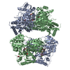
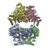
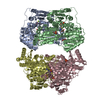
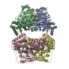
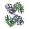
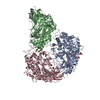
| Title | X-ray structure of phenylpyruvate decarboxylase in complex with 3-deaza-ThDP and 5-phenyl-2-oxo-valeric acid | ||||||
 Components Components | Phenylpyruvate decarboxylase | ||||||
 Keywords Keywords | LYASE / thiamine diphosphate / symmetrical dimer of dimers / closed active site loops / substrate complex | ||||||
| Function / homology |  Function and homology information Function and homology informationindolepyruvate decarboxylase / indolepyruvate decarboxylase activity / auxin biosynthetic process / : / pyruvate decarboxylase activity / thiamine pyrophosphate binding / magnesium ion binding / cytosol Similarity search - Function | ||||||
| Biological species |  Azospirillum brasilense (bacteria) Azospirillum brasilense (bacteria) | ||||||
| Method |  X-RAY DIFFRACTION / SYNCHROTRON / MOLECULAR REPLACEMENT / Resolution: 1.9 Å X-RAY DIFFRACTION / SYNCHROTRON / MOLECULAR REPLACEMENT / Resolution: 1.9 Å | ||||||
 Authors Authors | Versees, W. / Spaepen, S. / Wood, M.D. / Leeper, F.J. / Vanderleyden, J. / Steyaert, J. | ||||||
 Citation Citation | Journal: J.Biol.Chem. / Year: 2007 Title: Molecular mechanism of allosteric substrate activation in a thiamine diphosphate-dependent decarboxylase. Authors: Versees, W. / Spaepen, S. / Wood, M.D. / Leeper, F.J. / Vanderleyden, J. / Steyaert, J. | ||||||
| History |
| ||||||
| Remark 999 | SEQUENCE Authors state there is an error in the database sequence. The correct residue at position 327 is ARG. |
- Structure visualization
| Structure viewer | Molecule: MolmilJmol/JSmol |
|---|
- Downloads & links
-Download
| PDBx/mmCIF format | 2q5q.cif.gz | 243.8 KB | Display | PDBx/mmCIF format |
|---|---|---|---|---|
| PDB format | pdb2q5q.ent.gz | 189.3 KB | Display | PDB format |
| PDBx/mmJSON format | 2q5q.json.gz | Tree view | PDBx/mmJSON format | |
| Others |  Other downloads Other downloads |
-Validation report
| Arichive directory | https://data.pdbj.org/pub/pdb/validation_reports/q5/2q5qftp://data.pdbj.org/pub/pdb/validation_reports/q5/2q5q | HTTPS FTP |
|---|
-Related structure data
| Related structure data | 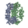 2q5jC 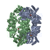 2q5lC  2q5oC 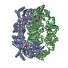 2nxwS S: Starting model for refinement C: citing same article ( |
|---|---|
| Similar structure data |


-Links
 PDBj
PDBj


- Assembly
| Deposited unit | 
| ||||||||||||
|---|---|---|---|---|---|---|---|---|---|---|---|---|---|
| 1 | 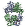
| ||||||||||||
| 2 |
| ||||||||||||
| Unit cell |
| ||||||||||||
| Components on special symmetry positions |
| ||||||||||||
| Details | The biological assembly is a tetramer generated from the dimer in the asymmetric unit by the operation: x,-y+1,-z |
-Components
-Protein , 1 types, 2 molecules AB
| #1: Protein | Mass: 60326.855 Da / Num. of mol.: 2 Source method: isolated from a genetically manipulated source Source: (gene. exp.) Azospirillum brasilense (bacteria) / Gene: ipdC / Plasmid: pET28a / Production host: |
|---|
-Non-polymers , 6 types, 1128 molecules 


| #2: Chemical |  Mass: 24.305 Da / Num. of mol.: 2 / Source method: obtained synthetically / Formula: Mg Mass: 24.305 Da / Num. of mol.: 2 / Source method: obtained synthetically / Formula: Mg#3: Chemical |  Mass: 423.318 Da / Num. of mol.: 2 / Source method: obtained synthetically / Formula: C13H19N3O7P2S Mass: 423.318 Da / Num. of mol.: 2 / Source method: obtained synthetically / Formula: C13H19N3O7P2S#4: Chemical | ChemComp-KPV /  Mass: 192.211 Da / Num. of mol.: 4 / Source method: obtained synthetically / Formula: C11H12O3 Mass: 192.211 Da / Num. of mol.: 4 / Source method: obtained synthetically / Formula: C11H12O3#5: Chemical | ChemComp-TLA / |  Mass: 150.087 Da / Num. of mol.: 1 / Source method: obtained synthetically / Formula: C4H6O6 Mass: 150.087 Da / Num. of mol.: 1 / Source method: obtained synthetically / Formula: C4H6O6#6: Chemical |  Mass: 92.094 Da / Num. of mol.: 2 / Source method: obtained synthetically / Formula: C3H8O3 Mass: 92.094 Da / Num. of mol.: 2 / Source method: obtained synthetically / Formula: C3H8O3#7: Water | ChemComp-HOH / | Mass: 18.015 Da / Num. of mol.: 1117 / Source method: isolated from a natural source / Formula: H2O |
|---|
-Experimental details
-Experiment
| Experiment | Method: X-RAY DIFFRACTION / Number of used crystals: 1 |
|---|
- Sample preparation
| Crystal | Density Matthews: 2.18 Å3/Da / Density % sol: 43.54 % |
|---|---|
| Crystal grow | Temperature: 293 K / Method: vapor diffusion, hanging drop / pH: 6.5 Details: 11% PEG3350, 0.2 M di-ammonium tartrate, pH 6.5, VAPOR DIFFUSION, HANGING DROP, temperature 293K |
-Data collection
| Diffraction | Mean temperature: 100 K |
|---|---|
| Diffraction source | Source: SYNCHROTRON / Site: EMBL/DESY, HAMBURG  / Beamline: BW7A / Wavelength: 0.9732 Å / Beamline: BW7A / Wavelength: 0.9732 Å |
| Detector | Type: MAR CCD 165 mm / Detector: CCD / Date: May 30, 2006 |
| Radiation | Monochromator: Fixed exit double crystal Si [111], horizontally focussing Protocol: SINGLE WAVELENGTH / Monochromatic (M) / Laue (L): M / Scattering type: x-ray |
| Radiation wavelength | Wavelength: 0.9732 Å / Relative weight: 1 |
| Reflection | Resolution: 1.9→34.9 Å / Num. all: 83208 / Num. obs: 76991 / % possible obs: 92.5 % / Observed criterion σ(I): -3 / Redundancy: 4.8 % / Biso Wilson estimate: 20.54 Å2 / Rsym value: 0.074 / Net I/σ(I): 18.4 |
| Reflection shell | Resolution: 1.9→1.97 Å / Redundancy: 2.5 % / Mean I/σ(I) obs: 2.45 / Num. unique all: 8220 / Rsym value: 0.48 / % possible all: 92 |
- Processing
| Software |
| |||||||||||||||||||||||||
|---|---|---|---|---|---|---|---|---|---|---|---|---|---|---|---|---|---|---|---|---|---|---|---|---|---|---|
| Refinement | Method to determine structure: MOLECULAR REPLACEMENT Starting model: PDB entry 2NXW Resolution: 1.9→34.9 Å / Cross valid method: THROUGHOUT / σ(F): 0 / σ(I): 0 / Stereochemistry target values: Engh & Huber
| |||||||||||||||||||||||||
| Refinement step | Cycle: LAST / Resolution: 1.9→34.9 Å
| |||||||||||||||||||||||||
| Refine LS restraints |
| |||||||||||||||||||||||||
| LS refinement shell | Resolution: 1.9→1.91 Å
|
